
Inquiry Basket
 Loading ......
Epstein-Barr virus was first successfully established by Epstein and Barr in 1964 from Burkitt African childhood lymphoma cells through in vitro suspension culture; in 1968, Henle et al. discovered that EBV was the causative factor of infectious mononucleosis; in 1970, it was identified as related to nasal and pharyngeal cancer; it was identified as related to the incidence of non-Hodgkin's lymphoma in 1980.
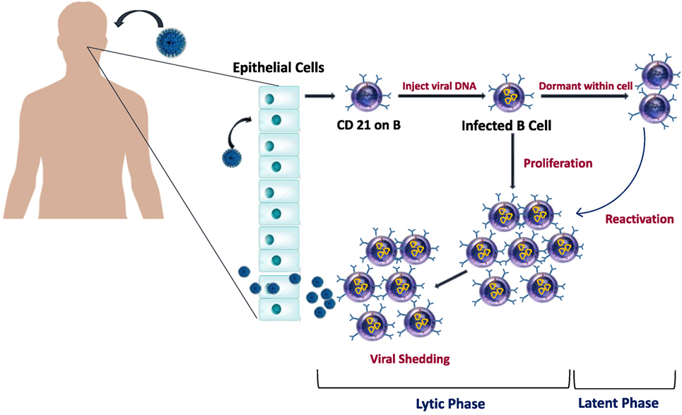 Figure 1. Epstein–Barr virus (EBV) life cycle in healthy carriers.(Source: Smatti MK, et al. 2018)
Epstein-Barr virus is also called human herpesvirus type 4. It is a DNA oncogenic virus of the genus Lymphotropic Virus of the γ subfamily. It mainly infects lymphocytes in the oropharynx. It can also infect epithelial cells and fibroblasts. The main route of transmission is oral close contact. It mostly occurs in childhood, most cases have a good prognosis, except for those with immune deficiencies who may experience life-threatening infections. EB virus is the most common form of latent infection. More than 90% of people have lifelong latent infection with EB virus. The virus is activated under certain conditions and causes cancer. Current research has found that EBV is associated with nasopharyngeal cancer, infectious mononucleosis, Hodgkin and non-Hodgkin lymphoma, African childhood lymphoma (Burkitt lymphoma), and epithelial cell carcinomas including gastric cancer.
The morphology of EB virus is similar to that of other herpes viruses, with a round shape and a diameter of 180 to 200 nm. The basic structure contains three parts: nucleoid, capsid and envelope. The nucleoid is a dense object with a diameter of 45 nm, mainly containing double-stranded linear DNA. Its length varies with different strains, with an average molecular weight of 17.5 × 10⁴ bp. The capsid has icosahedron stereosymmetry and is composed of 162 shell particles. The envelope consists of the nuclear membrane of infected cells, which contains virus-encoded membrane glycoproteins, which have functions such as recognizing EBV receptors on lymphocytes and fusion with cells. In addition, there is a protein coat between the capsule and capsid.
EBV infection is sporadic and widely distributed in the population. Among them, the sources of infection are virus carriers and patients. Close oral contact is the main mode of transmission, but it can also be transmitted through blood transfusion.
The initial site of EBV replication is the oropharynx, where it grows and reproduces in B lymphocytes and oral epithelial cells, and then infects B lymphocytes. These cells enter the blood circulation in large numbers and cause systemic infection. And it can lie latent in human lymphoid tissue for a long time. When the body's immune function is low, the latent EB virus is activated to form a recurrent infection. It is generally believed that cellular immunity (such as the cytotoxic response of T lymphocytes) plays a key role in "monitoring" viral activation and clearing transformed B lymphocytes.
The incubation period of IM is 5 to 15 days, usually 9 to 11 days; the onset varies, and about 40% of patients have prodromal symptoms, which last for 4 to 5 days, such as fatigue, headache, anorexia, nausea, loose stools, and chills etc.; although the symptoms of this disease are diverse, most of them can have more typical symptoms. It mainly affects children and adolescents and is clinically characterized by: fever, sore throat, enlarged liver, spleen and lymph nodes, increased lymphocytes and atypical lymphocytes in peripheral blood.
After the human body is infected with EB virus, it can produce membrane shell antibodies, anti-membrane antibodies, early antibodies, neutralizing antibodies, complement fixation antibodies, virus-related nuclear antibodies, etc. Among them, anti-VCAIgM and IgG are more commonly used. The former appears early, disappears quickly, has high sensitivity and specificity, and has early diagnostic value. The latter appears early, has a higher titer and lasts for life, and is suitable for study investigationin epidemic diseases.
Peripheral blood EBV-DNA is a method with high sensitivity and specificity, which can achieve the purpose of early diagnosis when the antibody response is not yet clear. However, at the 14th day of the disease course, viral nucleic acid was no longer detectable in the serum of most EBV-IM patients, and after the 22nd day of the disease course, no EBV nucleic acid was detected in the serum of EBV-IM patients.
EBV viral load testing: EBV viral load testing can differentiate between low-level replication in healthy EBV carriers and high-level active infection in patients with EBV-related diseases. Patients with active EBV infection or EBV-related tumors often have high levels of EBV-DNA loads in their serum or plasma, while healthy EBV carriers may have low-level EBV-DNA loads in their blood lymphocytes, which cannot be detected EBV-DNA in their serum or plasma.
Reference
Loading ......
