
Inquiry Basket
 Loading ......
Autoantigens are self-derived molecules that are mistakenly recognized by the immune system as foreign targets. Under normal physiological conditions, the immune system maintains tolerance to the body's own proteins, nucleic acids, lipids, carbohydrates, and cellular structures. In autoimmune diseases, this immune tolerance is disrupted. As a result, autoreactive B cells and T cells may respond to self-antigens and produce autoantibodies or cell-mediated immune responses that contribute to tissue inflammation, organ damage, and disease progression.
Autoantigens can be intracellular, cell-surface, secreted, tissue-specific, or widely expressed molecules. Many clinically important autoantigens are proteins or protein complexes involved in essential biological functions such as DNA replication, RNA processing, intracellular signaling, enzyme catalysis, structural organization, coagulation, metabolism, and immune regulation. Examples include myeloperoxidase (MPO) and proteinase 3 (PR3) in ANCA-associated vasculitis, Ro/SSA and La/SSB in systemic autoimmune diseases, tissue transglutaminase (tTG) in celiac disease, thyroglobulin and thyroid peroxidase in autoimmune thyroid disease, and insulin or glutamic acid decarboxylase 65 (GAD65) in autoimmune diabetes.
The clinical importance of autoantigens is closely linked to the autoantibodies that recognize them. Autoantibody profiles can provide valuable information for disease screening, differential diagnosis, classification, risk stratification, prognosis, and monitoring. In many cases, disease-associated autoantibodies appear before obvious clinical symptoms, making autoantigen-based immunoassays useful tools for early detection and disease research.
Autoimmune diseases are highly heterogeneous. Some are organ-specific, such as autoimmune thyroid disease, type 1 diabetes, autoimmune gastritis, and celiac disease. Others are systemic, such as systemic lupus erythematosus, rheumatoid arthritis, antiphospholipid syndrome, Sjögren's syndrome, systemic sclerosis, idiopathic inflammatory myopathies, and ANCA-associated vasculitis. In each disease area, autoantigens provide the molecular basis for detecting disease-related autoantibodies.
For example, in ANCA-associated vasculitis, MPO and PR3 are the two major target antigens for anti-neutrophil cytoplasmic antibodies. In celiac disease, tTG and deamidated gliadin peptides are important autoantigenic targets. In antiphospholipid syndrome, β2-glycoprotein I and cardiolipin-associated antigenic complexes are central to laboratory investigation. In myositis, autoantigens such as Mi-2, Jo-1, PL-7, PL-12, SRP54, MDA5, TIF1-γ, NXP2, PM/Scl, and Ku support the detection of myositis-specific and myositis-associated autoantibodies. In autoimmune liver diseases, antigens such as PDC-E2, gp210, sp100, SLA/LP, and liver cytosol antigen 1 are widely used in diagnostic and research assays.
Autoantigens used in IVD assays may be produced as recombinant proteins, purified native proteins, synthetic peptides, fusion proteins, protein fragments, or antigen complexes. Each format has advantages and limitations. The best option depends on the disease target, relevant epitopes, assay format, and manufacturing requirements.
Recombinant autoantigens are widely used because they offer defined sequence composition, scalable production, and controlled manufacturing. Expression systems such as E. coli, yeast, insect cells, and mammalian cells can be selected according to the structural requirements of the antigen. E. coli expression is often suitable for non-glycosylated proteins, linear epitopes, enzyme fragments, and cost-effective production. Yeast systems may provide improved folding for some eukaryotic proteins and scalable fermentation. Insect cells and mammalian cells are often preferred for complex proteins requiring disulfide bonds, glycosylation, secretion, or native-like folding.
Native autoantigens are purified from natural biological sources and may retain native post-translational modifications or conformational structures. They can be valuable when recombinant versions fail to reproduce clinically relevant epitopes. However, native antigens may present challenges related to source availability, purification complexity, biological variability, and scalability.
Synthetic peptides are useful when autoantibody recognition depends on a defined linear epitope. They offer high consistency and simplified production, but they may not capture conformational epitopes. Fusion proteins and chimeric constructs can improve solubility, expression yield, immobilization, and epitope presentation. For some disease markers, antigen fragments may perform better than full-length proteins because they expose immunodominant regions while reducing nonspecific background.
Selecting the right expression system is one of the most important decisions in autoantigen development. The choice affects yield, solubility, folding, post-translational modifications, purification strategy, cost, and assay performance. No single expression platform is ideal for every autoantigen.
For antigens dominated by linear epitopes, bacterial expression can be efficient and cost-effective. Many autoantigens used in line blot and ELISA formats can be produced in E. coli, especially when the antigen does not require glycosylation or complex folding. However, bacterial systems may generate inclusion bodies, lack eukaryotic modifications, and sometimes produce proteins with reduced conformational antigenicity.
Yeast expression systems offer advantages for secreted or moderately complex proteins. They can support disulfide bond formation and scalable fermentation. However, yeast-specific glycosylation patterns may differ from mammalian glycosylation and should be evaluated if glycan-dependent epitopes are relevant. Insect cell expression is useful for eukaryotic proteins requiring better folding and post-translational processing than bacterial systems can provide. Mammalian expression is often selected for complex glycoproteins, membrane-associated extracellular domains, and antigens where native-like folding is essential.
In many projects, multiple expression strategies may be screened before selecting the final antigen. Analytical evaluation should be paired with functional testing using clinically relevant serum or plasma panels. A high-yield antigen is not necessarily the best diagnostic antigen if it fails to capture the intended autoantibody population.
Creative Diagnostics provides autoantigens covering major autoimmune disease areas and assay development needs. These products are designed to support researchers, diagnostic developers, and manufacturers working on autoantibody detection platforms. The portfolio includes antigens associated with systemic autoimmune diseases, organ-specific autoimmune diseases, inflammatory disorders, neurological autoimmune syndromes, and disease-specific diagnostic panels.
Table 1. Representative Autoantigen Categories
| Product Classification | Representative Autoantigens | IVD and Research Relevance |
| ANCA Associated Diseases | MPO, PR3 | Detection of ANCA-related autoantibodies in vasculitis research and immunoassay development |
| Antinuclear Antibodies (ANA) | dsDNA, Sm, RNP, Ro/SSA, La/SSB, Scl-70, centromere proteins | Supports systemic autoimmune disease panels and ANA profile assays |
| Antiphospholipid Syndrome | β2-glycoprotein I, cardiolipin-related targets, prothrombin | Used for antiphospholipid antibody assay development |
| Autoimmune Hepatitis and Liver Disease | SLA/LP, LC1, PDC-E2, gp210, sp100 | Supports autoimmune liver disease marker panels |
| Autoimmune Thyroid Disease | Thyroglobulin, thyroid peroxidase, TSH receptor | Used in thyroid autoantibody testing workflows |
| Celiac Disease | Tissue transglutaminase, deamidated gliadin peptides | Supports celiac disease screening and confirmatory assays |
| Crohn's Disease and IBD | ASCA-related antigens, pancreatic targets, microbial-associated antigens | Supports inflammatory bowel disease research panels |
| Dermatology | DSG1, DSG3, BP180, BP230, collagen VII | Relevant for autoimmune blistering disease assays |
| Diabetes Mellitus | Insulin, GAD65, IA-2, ZnT8 | Supports autoimmune diabetes risk and marker studies |
| Membranous Nephropathy | PLA2R, THSD7A | Used in membranous nephropathy autoantibody detection |
| Myositis | Jo-1, PL-7, PL-12, Mi-2, MDA5, TIF1-γ, NXP2, SRP54, PM/Scl, Ku | Supports myositis-specific and myositis-associated antibody panels |
| Primary Biliary Cholangitis (PBC) | PDC-E2, BCOADC-E2, OGDC-E2, gp210, sp100, PML protein, Centromere proteins, KLHL12 | Used in hepatic autoimmunity diagnostics and cholestatic biomarker research |
| Paraneoplastic Syndrome | Hu, Yo, Ri, amphiphysin, CV2/CRMP5, Ma2, recoverin | Used in neurological autoimmunity and paraneoplastic antibody research |
| Rheumatoid Arthritis | Citrullinated peptides, rheumatoid factor-related targets, RA33 | Supports rheumatoid arthritis immunoassay development |
A successful autoantigen reagent begins with careful design. The design strategy should reflect the biological characteristics of the target and the intended assay format. For some targets, the full-length protein provides the best epitope coverage. For others, a specific domain or epitope-rich fragment provides better performance. In some cases, removal of hydrophobic regions, transmembrane domains, low-complexity regions, or unstable sequences can improve solubility and assay behavior.
Tags may be added to facilitate purification or immobilization. Common tags include His-tag, GST, MBP, Fc, biotin acceptor sequences, and other affinity handles. However, tags can influence antigen folding, solubility, background reactivity, or immobilization orientation. For IVD applications, tag placement and tag removal should be evaluated carefully. When possible, functional assay testing should compare different constructs to determine which format provides the best clinical discrimination.
Post-translational modifications are another important consideration. Some autoantibody epitopes depend on phosphorylation, citrullination, glycosylation, deamidation, oxidation, or conformational structure. In rheumatoid arthritis, citrullinated peptides and proteins are central to anti-citrullinated protein antibody detection. In celiac disease, deamidated gliadin peptides improve disease-related antibody recognition. For glycoprotein antigens, expression host selection may influence glycosylation and assay performance.
Quality control for autoantigens should combine biochemical characterization and functional immunological testing. Biochemical assays confirm identity, purity, concentration, molecular weight, aggregation state, and stability. Functional assays confirm whether the antigen binds expected autoantibodies and avoids nonspecific binding with negative or unrelated disease samples.
Recommended QC tests may include SDS-PAGE, Western blot, protein concentration analysis, endotoxin testing when relevant, HPLC or SEC-HPLC, mass spectrometry, residual host-cell protein assessment, residual DNA testing, and immunoreactivity testing with reference sera or characterized monoclonal antibodies. Additional studies may include accelerated stability, real-time stability, freeze-thaw stability, shipping stress, coating stability, conjugation compatibility, and lot-to-lot comparison.
Assay validation should include sensitivity, specificity, precision, reproducibility, linearity when applicable, interference, cross-reactivity, cut-off determination, and comparison against reference methods or predicate devices. Autoantigen quality directly affects all of these performance characteristics.
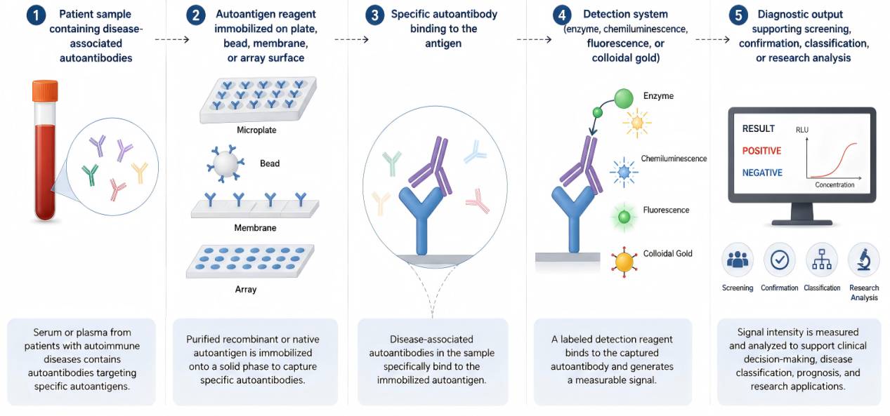 Figure 1. Role of Autoantigens in Autoimmune IVD Assay Development
Autoantigen-based assay development typically follows a structured workflow. The process begins with target selection based on disease relevance, literature support, clinical need, and assay format. Next, antigen design is performed to determine whether the final reagent should be full-length, truncated, modified, native, recombinant, peptide-based, or complex-based. Expression and purification are then optimized to obtain sufficient yield and purity. After biochemical characterization, the antigen is tested in the intended assay format using clinical or reference samples.
Optimization may involve coating concentration, immobilization chemistry, blocking reagent, sample dilution, wash buffer, conjugate selection, incubation time, and detection substrate. Once the assay conditions are established, validation studies are conducted to evaluate analytical and clinical performance. For commercial IVD development, the antigen manufacturing process must be controlled to support reproducible production, documentation, and quality release criteria.
Autoantigen-based diagnostic development can be technically challenging. One major challenge is epitope preservation. Autoantibodies may recognize conformational epitopes that are sensitive to denaturation, immobilization, or chemical coupling. Another challenge is antigen heterogeneity. Some autoantigens exist as isoforms, splice variants, modified proteins, or multi-protein complexes. Selecting the wrong antigen form may reduce clinical sensitivity.
Background reactivity is also a common issue. Human samples contain many proteins, antibodies, rheumatoid factor, heterophilic antibodies, and matrix components that can cause nonspecific signal. Antigen impurities, exposed tags, aggregates, and inappropriate blocking conditions can further increase background. For multiplex assays, cross-reactivity between antigens or detection channels must be carefully controlled.
Manufacturing consistency is another important factor. An antigen that performs well in a small pilot batch must also be reproducible at larger scale. Changes in expression host, fermentation conditions, purification resin, buffer composition, storage temperature, or lyophilization conditions may affect antigen performance. Therefore, IVD developers should evaluate antigen lots under conditions that reflect the final assay workflow.
Autoantigens serve as the critical molecular interface between patient-derived autoantibodies and detection systems, directly influencing assay sensitivity, specificity, reproducibility, and overall clinical performance. Consequently, careful consideration of antigen selection, expression system, purification strategy, and validation approach is required to ensure compatibility with the intended diagnostic platform.
With the continuous advancement of autoimmune diagnostics toward higher sensitivity, automation, multiplexing, and disease-specific testing panels, the demand for reliable and well-characterized autoantigens continues to grow. High-quality recombinant and native autoantigens enable robust immunoassay development and facilitate accurate investigation of disease mechanisms, biomarker identification, and immune response profiling.
The Creative Diagnostics Autoantigen Portfolio is developed to address these evolving needs. It provides a diverse range of high-quality reagents, including recombinant proteins, native antigens, antigen fragments, and synthetic peptides. These products support a wide spectrum of applications such as assay development, antibody characterization, calibrator and control preparation, and diagnostic research.
Designed with assay compatibility in mind, the portfolio supports multiple IVD platforms, including ELISA, chemiluminescent immunoassays (CLIA), line immunoassays, immunoblotting, lateral flow assays, bead-based systems, and microarray technologies. With broad disease coverage—spanning ANCA-associated diseases, antinuclear antibody (ANA) profiles, antiphospholipid syndrome, autoimmune liver and thyroid diseases, celiac disease, Crohn's disease, dermatological autoimmune conditions, diabetes mellitus, membranous nephropathy, myositis, paraneoplastic syndromes, and rheumatoid arthritis—the portfolio enables both single-analyte testing and multiplex panel development.
In addition to standard catalog products, Creative Diagnostics offers custom autoantigen development services. These services support specialized project requirements, including construct design, expression optimization, protein modification, labeling, conjugation, formulation, and assay-specific validation. This flexibility allows seamless progression from early-stage biomarker discovery to fully developed commercial IVD applications.
References
Loading ......
