
Inquiry Basket
 Loading ......
Multiplex Immunohistochemistry (mIHC) is a cutting-edge fluorescence-based detection technology that combines advanced immunohistochemistry with multi-channel labeling. This innovative approach enables comprehensive spatial and phenotypic analysis of the tumor immune microenvironment within a single tissue section. By simultaneously detecting multiple biomarkers, researchers can quantitatively assess cellular abundance, functional states, and spatial relationships, providing unprecedented insights into cellular interactions and microenvironment dynamics. With superior sensitivity, specificity, and tissue structure preservation, Magic-Fluor™ mIHC is a powerful tool for advancing research and diagnostics in oncology and immunology.
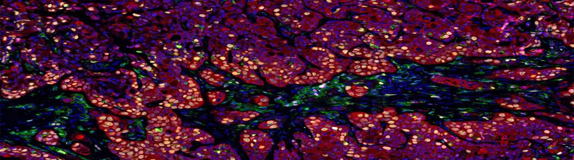 Fig. 1. Multiplex Immunohistochemistry
Magic-Fluor™ mIHC (Multiplex Immunohistochemistry, mIHC) employs Tyramide Signal Amplification (TSA) technology, an enzymatic detection system utilizing horseradish peroxidase (HRP)-conjugated secondary antibodies. Upon binding to primary antibodies, HRP catalyzes the conversion of fluorescent tyramide substrates into reactive intermediates. These intermediates covalently bind tyrosine residues near the target antigen, depositing stable fluorescent signals directly onto the sample. After completing this step, heat treatment is used to remove unbound antibodies to eliminate background noise and improve signal specificity. Subsequently, by replacing with another specific primary antibody (primary antibody) and using different fluorescent substrates, a second round of incubation is carried out. This iterative process allows simultaneous visualization of multiple targets on a single sample, ensuring high-resolution, multiplexed imaging with minimal background noise.
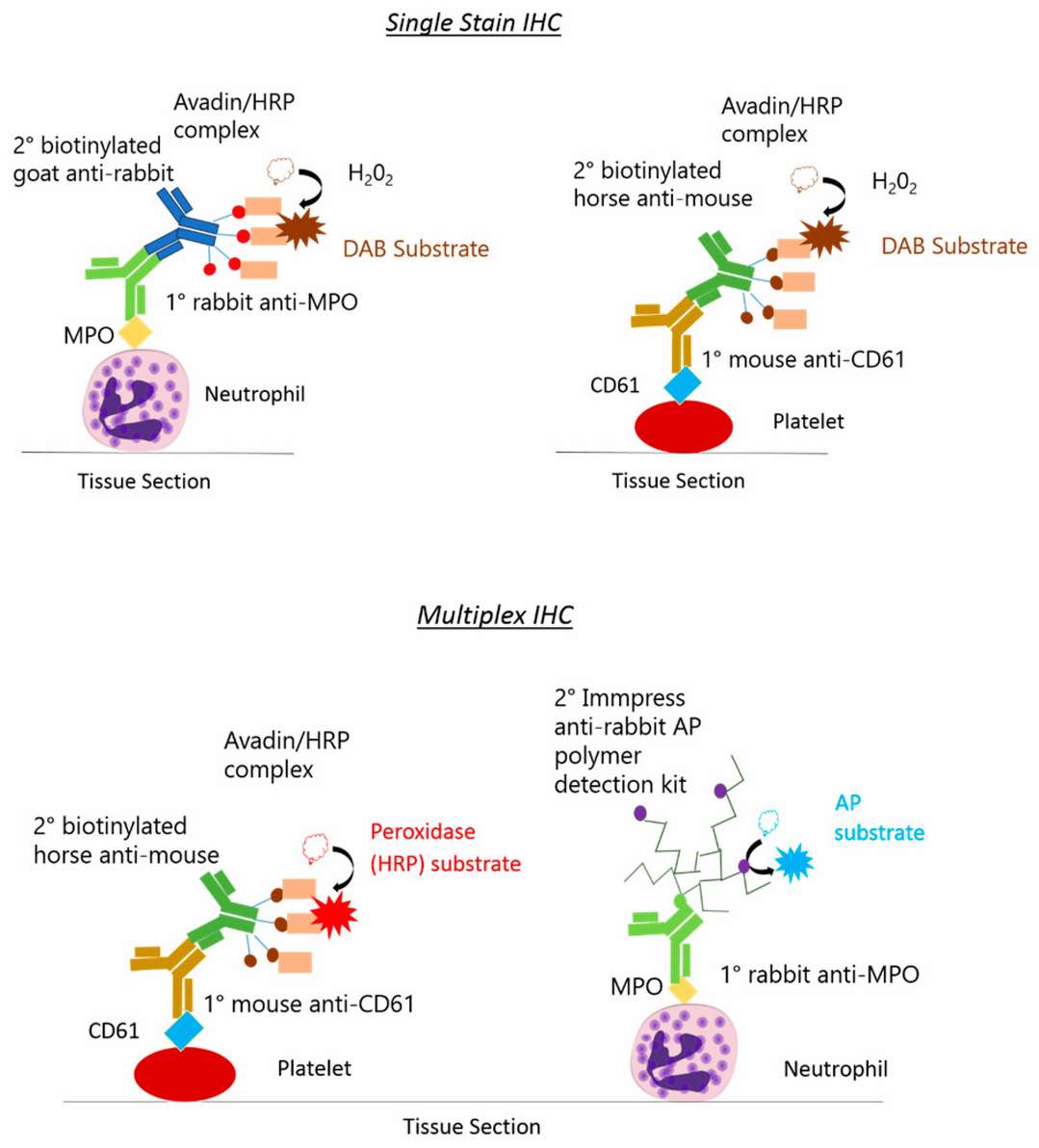 Fig. 2. Single stain IHC and Multiplex IHC
Tertiary lymphoid structures (TLS) are organized aggregates of immune cells that form postnatally in non-lymphoid tissues under pathological conditions such as autoimmune diseases, chronic infections, and solid tumors. Composed of B cells, T cells, dendritic cells, macrophages, stromal cells, and endothelial cells, TLS mimic the functional properties of secondary lymphoid organs, fostering localized immune activation.
Within the tumor microenvironment (TME), TLS enhance antitumor immunity by recruiting and organizing immune cells into solid tumors, contributing to effective immune surveillance and cytotoxic responses. Their presence is strongly correlated with improved clinical outcomes across multiple cancer types, highlighting their potential as biomarkers for favorable prognosis and as therapeutic targets.
This dynamic interplay between TLS and the TME underscores their significance in bridging innate and adaptive immunity, offering promising avenues for advancing cancer immunotherapy strategies.
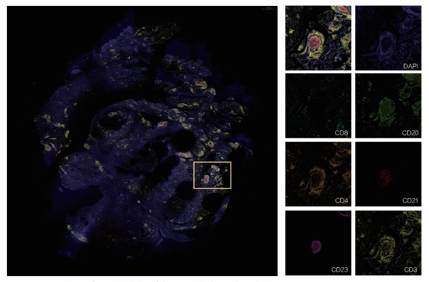 Fig. 3. Breast cancer tissue (6 markers 7-colors mIHC).
* The yellow box is a local magnification image (shown on the right).
Through in situ tissue analysis enables automatic recognition of tertiary lymphoid structures (TLS) and precise, single-cell quantitative analysis of the spatial distribution of surrounding immune cells. This approach allows for detailed mapping of immune cell interactions and spatial dynamics within the tissue microenvironment.
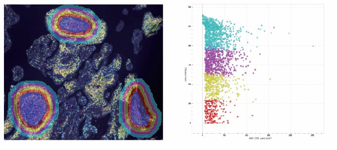 Fig. 4. Different colors represent specific distance ranges from tertiary lymphoid structures (TLS). In the scatter plot, these colors indicate the number of CD8⁺ cells at corresponding distances from TLS.
Through advanced tumor-specific staining, our reagents automatically recognizes tumor and stromal areas, enabling micrometer-precision single-cell spatial quantitative analysis of cells at the tumor boundary. This approach generates comprehensive quantified data on the expression levels of multiple positive cells within the tumor microenvironment and the interaction distance patterns among different phenotypic cells.
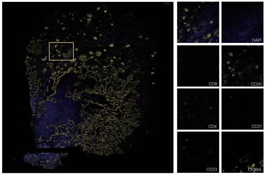 Fig. 5. Human squamous cell carcinoma tissue (6 markers 7-colors mIHC)
* The yellow box is a local magnification image on the right.
Centering on the tumor, we perform micrometer-precision, single-cell quantification analyses of the spatial distribution of immune cells within the surrounding microenvironment.
 Fig. 6. Different colors represent different distance ranges from the tumor. In the scatter plot, different colors indicate the number of CD8+ cells at different distances from the tumor.
Macrophages are essential immune cells involved in maintaining homeostasis, repairing tissue damage, and combating pathogens. Tumor-associated macrophages (TAMs) can constitute up to 50% of the cellular content in some solid tumors and play crucial roles in tumor initiation, progression, and metastasis. Within the tumor microenvironment (TME), TAMs polarize into M1 or M2 phenotypes, each expressing specific marker genes and performing distinct functions. M1-type TAMs exhibit high levels of aerobic glycolysis and produce reactive oxygen species (ROS) to eliminate pathogens and mediate anti-tumor responses. In contrast, M2-type TAMs rely on elevated oxidative phosphorylation and promote the growth of malignant tumor cells.
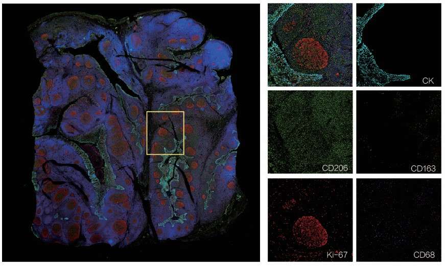 Fig. 7. Tonsil (5 markers 6-colors mIHC staining).
* The yellow box is a local magnification image on the right.
Using in situ single-cell scatter plot and t-SNE image analysis functions, we identify cells and their subcellular localization through multichannel protein staining, projecting cellular characteristics-such as staining intensity, area, and morphology-onto a two-dimensional scatter plot. This enables precise single-cell quantitative analyses, including phenotype characterization and co-localization assessments.
 Fig. 8. Precision quantification analysis
Neovascularization is crucial for tumor growth and progression. Tumors larger than 1–2 mm initiate angiogenesis to obtain oxygen and nutrients, facilitating immune suppression and evasion. Cancer cells can migrate along blood vessel surfaces, integrating into the developing tumor.
Cancer-associated fibroblasts (CAFs), transformed from normal fibroblasts under tumor influence, are key heterogeneous components of the tumor microenvironment. CAFs secrete specific cytokines and extracellular matrix (ECM) components that regulate tumor cell proliferation, metastasis, drug resistance, and other malignant behaviors.
The ECM-composed of proteoglycans, glycoproteins, collagen, and elastin-promotes cell communication by serving as a substrate for cytokines, growth factors, and chemokines. It facilitates cell adhesion and migration, forming complex signaling networks within the tumor.
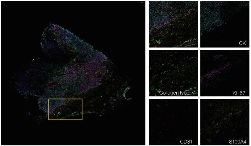 Fig. 9. Human colorectal cancer tissue(5 markers 6-colors mIHC).
* The yellow box is a local magnification image on the right.
Loading ......
