
Inquiry Basket
 Loading ......
Cervical cancer is a malignant tumor that seriously endangers women's health, and its incidence ranks second among female malignant tumors in the world. The occurrence of cervical cancer is a continuous process, that is, from disordered cell differentiation to dysplasia, to carcinoma in situ, and finally to cervical cancer. Many studies have found that persistent infection with human papillomavirus (HPV) is closely related to the evolution of cervical lesions. HPV can promote the occurrence of intrauterine epithelioid neoplasia and cervical cancer. The 25th International Papillomavirus Conference held in May 2009 clarified that the global HPV infection rate is on the rise and new genotypes have emerged. As a double-stranded circular DNA virus, HPV is divided into high-risk and low-risk types based on its biological characteristics and carcinogenic potential. The low-risk type mainly causes exophytic condyloma-like lesions, flat condyloma-like lesions, and cervical intraepithelial neoplasia (CIN) in the perianal skin of the genital tract and lower part of the vagina. The probability of inducing cervical cancer is less than 5%; the high-risk type causes CIN Ⅱ, CIN Ⅲ and The probability of cervical cancer is greater than 90%. HPV16, 18, 31, 33, 35, 39, 45, 51, 52, 56, 58, 59, and 68 are high-risk types and are usually detected in severe atypical hyperplasia and cervical cancer. The two proteins encoded by E6 and E7 in its structure can promote the transformation of cells from normal to malignant transformation. The proto-oncoproteins encoded by E6 and E7 genes are important causes of cervical cancer. Most HPV infections are transient, and only persistent HPV infections can lead to cervical cancer. Single or multiple infections of multiple types of HPV often exist in female cervical lesions. HPV testing is valuable for excluding CIN during routine cervical cancer screening and can distinguish high-risk women during follow-up. If the above screening methods are used properly, more than 98% of early-stage patients can be detected. It can be seen that the detection of HPV is of great significance for the diagnosis and prevention of cervical cancer. Currently, a variety of experimental projects have been carried out in clinical and laboratory settings to detect and screen HPV. The progress of HPV detection methods is summarized as follows.
Since HPV cannot proliferate in vitro, the morphological method originated in the 1940s is a traditional method, which mainly includes Pap smear cytopathological detection, electron microscopy technology (direct observation of viral particles), etc.
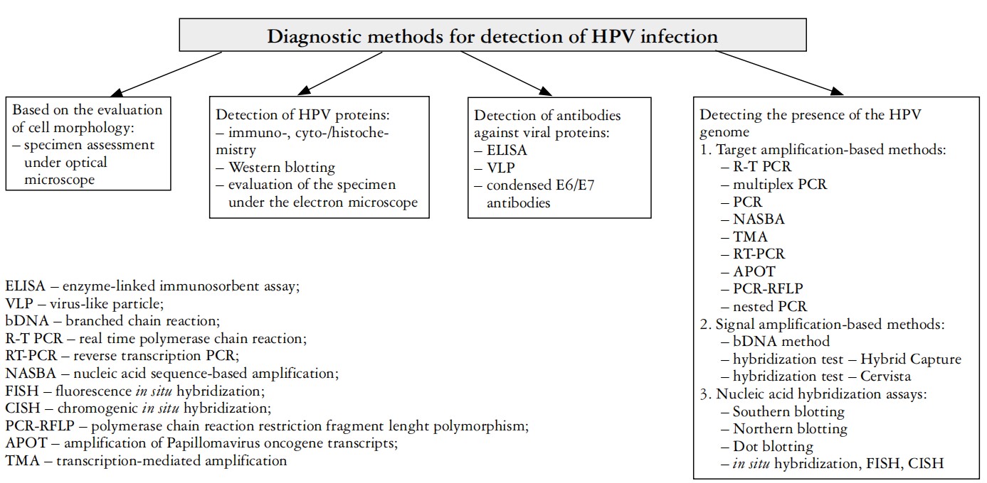 Figure 1. Diagnostic methods for detection of HPV infection.(Bogusiak K, et al., 2014)
Hollow cells are the main morphological changes of HPV infection. The diagnosis can be made if the hollow cells caused by HPV can be seen by Pap staining in the cervical transition zone. There are many factors that affect Pap smears, and the false negative rate is high, which limits its application. In recent years, with the advancement of specimen collection, production, and reading technology, traditional Pap smears have developed into liquid-based thin-layer cell technology (TCT) and automatic smear detection systems. TCT has relatively small impact, The operation is standardized, and its detection accuracy is higher than that of conventional Pap smear, and it is used clinically for large-scale census and screening. Since other viral infections and human factors may cause vacuoles or hollow changes in cells, as well as the influence of factors such as material sampling, staining, and subjective judgment, morphological detection of HPV still has low sensitivity, poor specificity, and false positives. It has the disadvantages of high negative rate and false positive rate, and it cannot classify HPV.
Types of colposcopes with electron microscopy technology include: traditional optical colposcope, photoelectric integrated colposcope and electronic colposcope. HPV is one of the earliest viruses to be observed under an electron microscope. The biopsy specimen is negatively stained. If characteristic virus particles can be seen under an electron microscope, a diagnosis can be made. Although electron microscopy is highly accurate, it is time-consuming, expensive instruments and equipment, and has a low positive rate. The specificity and sensitivity of electron microscopy for detecting HPV are not high, and it is not convenient to classify HPV, so it is currently rarely used.
In the 1950s, with the development of immunology, some antibodies could also be detected in serum and used as markers for the diagnosis and treatment of cervical cancer. Use recombinant technology to express antigens to detect corresponding antibodies in patient serum, or use antigen-immunized animals to prepare immune serum or monoclonal antibodies to detect HPV antigens in tissues or local mucus. The immunoadsorption method is commonly used clinically to detect the IgM and IgG antibodies of L1 virus-like particles in HPV and the E6 and E7 specific antibody proteins of HPV. The radioimmunoassay method is used to determine the levels of HPV16 antibodies in the serum of CIN and cervical cancer patients. In addition, immunohistochemistry is also used to detect HPV16 and 18 early protein E6 monoclonal antibodies, which can effectively prove the presence of E6 protein in infected tissues. The principle of this method is clear and the operation is relatively simple. However, since the objects of serological detection are antigens and antibodies, the human body has There is a certain delay in the immune response produced by HPV. At the same time, the HPV virus cannot be cultured and proliferated in vitro. Therefore, serological testing will miss detection in people without immune response and those infected with HPV latent period. In addition, most people may develop antibodies in their serum after being infected with HPV. Antibodies last for a long time in the body, some for several years, and are not unique to cervical cancer. Therefore, HPV serological antibody detection is used for the diagnosis of cervical cancer. It is not specific and its clinical application is limited.
Polymerase chain reaction (PCR) detects HPV-DNA. The most critical factor in evaluating HPV detection methods is detection sensitivity. Detection of HPV DNA based on gene amplification is currently the most sensitive detection method. PCR can not only be used to detect viral load quantification, DNA sequencing and mutation analysis, but can also perform multiplex amplification and analyze multiple DNA sequences simultaneously, and PCR has the characteristics of relatively simple operation, time saving, and unrestricted specimen sources. Therefore, PCR Testing is currently the best method for HPV-DNA testing and typing. Its disadvantage lies in its high sensitivity, which can easily lead to false positive detection due to cross-contamination of samples. The operation of PCR typing is relatively cumbersome, and the number of samples tested at one time is limited, which limits its application in large-scale screening.
Dot hybridization or in situ hybridization is used to detect HPV-DNA based on different specimens, which has good specificity and sensitivity. Nucleic acid hybridization detection methods mainly include in situ hybridization (ISH), nucleic acid blotting, hybridization capture (HC), etc. ISH uses HPV probes to hybridize to HPV-DNA in tissues. Its advantage is beneficial to pathological analysis, but the stability of the hybrid chain is not high. During the hybridization process, some probes may be single-stranded, reducing the hybridization rate and affecting the detection rate of HPV-DNA. Nucleic acid blotting is suitable for HPV typing and HPV-DNA relative molecular mass identification. It was used in early research on HPV, but it requires fresh tissue specimens and is complicated to operate. HC is a new method in the 21st century. Its basic principle is to use efficient liquid-phase RNA-DNA hybridization method to capture HPV-DNA in the sample. An alkaline phosphatase-labeled anti-RNA-DNA antibody-chemiluminescent signal display system was used.
HC-I can detect 9 high-risk HPV types, but its detection sensitivity is lower than PCR and other gene amplification technologies, and its clinical application is limited. Currently, HC-II is widely used to detect viral DNA and has a high negative predictive value for cervical lesions and cervical cancer. This method has been recognized worldwide and is widely used for screening and review of cervical cancer.
Gene chip technology is a new technology that was developed at the end of the 20th century. Its principle is to use in-situ synthesis or direct spotting methods to arrange a large number of DNA fragments or oligosaccharide nucleotide fragments on the surface of a support to produce two DNA fragments. The DNA probe array is then hybridized with the labeled sample, and the quantity and sequence information of the sample molecules are obtained by detecting the intensity of the hybridization signal, so as to study the gene sequence and function. The gene chip method has the advantages of miniaturization, intensification and standardization, and high throughput. The sensitivity and specificity of gene chips are higher than other methods. It can not only be used for typing, but also detect multiple types of mixed infections at the same time. However, due to the high cost of detection, it is difficult to put it into practical clinical application. However, with the development of this technology, if the cost decreases, it will undoubtedly be the most potential detection and analysis method in the future.
Colloidal gold technology is a new technology developed in recent years. It is a solid-phase labeled immunoassay technology that organically combines various methods such as colloidal gold labeling immunoassay technology and chromatography analysis technology. Immunocolloidal gold rapid diagnostic technology has the advantages of easy operation, easy determination, and rapidity. When testing samples, it only takes 5 to 10 minutes to determine the results, so it can achieve the purpose of rapid diagnosis. Colloidal gold is cheaper, faster, and easier to operate than HC-II, which is widely used at present. It can be used as a screening test for cervical cancer. It is undoubtedly the future development direction of HPV detection.
Reference
| Target | Cat. No. | Product Name | Size | Species Reactivity | Application | Detection Sample | |
| HPV | DEIA-F678S | Human Papilloma Virus IgG ELISA kit | 96T | Human | Qualitative | plasma, serum | Inquiry |
| DEIASL404 | Human HPV18 IgM ELISA kit | 96T | Qualitative | Serum, plasma | Inquiry | ||
| DEIASL405 | Human HPV 16 L1-capsids IgG ELISA Kit | 96T | Qualitative | Serum, plasma | Inquiry | ||
| DEIASL406 | Human HPV 16 IgM ELISA Kit | 96T | Qualitative | Serum, plasma | Inquiry | ||
| DEIASL407 | Human Papilloma Virus IgM ELISA Kit | 96T | Qualitative | Serum, plasma | Inquiry | ||
| DEIASL408 | Human HPV 18 L1-capsids IgG ELISA Kit | 96T | Qualitative | Serum, plasma | Inquiry | ||
| DEIASL409 | Rabbit HPV 16 L1-capsids IgG ELISA Kit | 96T | Qualitative | Serum, plasma | Inquiry | ||
| DEIASL410 | Human high risk HPV L1-capsids (HR-HPVL1) IgG ELISA kit | 96T | Qualitative | Serum, plasma | Inquiry | ||
| DEIASL119 | HPV(18) Antigen ELISA Quantitation Kit | 96T | Quantitative | Serum, plasma | Inquiry | ||
| DEIASL120 | HPV(52) Antigen ELISA Quantitation Kit | 96T | Quantitative | Serum, plasma | Inquiry | ||
| DEIASL121 | HPV(58) Antigen ELISA Quantitation Kit | 96T | Quantitative | Serum, plasma | Inquiry | ||
| DEIASL171 | Human HPV 16 E7 Oncoprotein ELISA Kit | 96T | Human | Quantitative | Cell lysates, tissue lysates, cervical smears, plasma, serum | Inquiry | |
| DEIASL172 | Human HPV 18 E7 Oncoprotein ELISA Kit | 96T | Quantitative | Cell lysates, tissue lysates, or cervical smears | Inquiry | ||
| HPV16 | DEIASL118 | HPV (16) Antigen ELISA Quantitation Kit | 96T | Human | Quantitative | Serum and plasma | Inquiry |
Loading ......
