






 Autoimmunity Catalogue
Inquiry Basket
 Loading ......
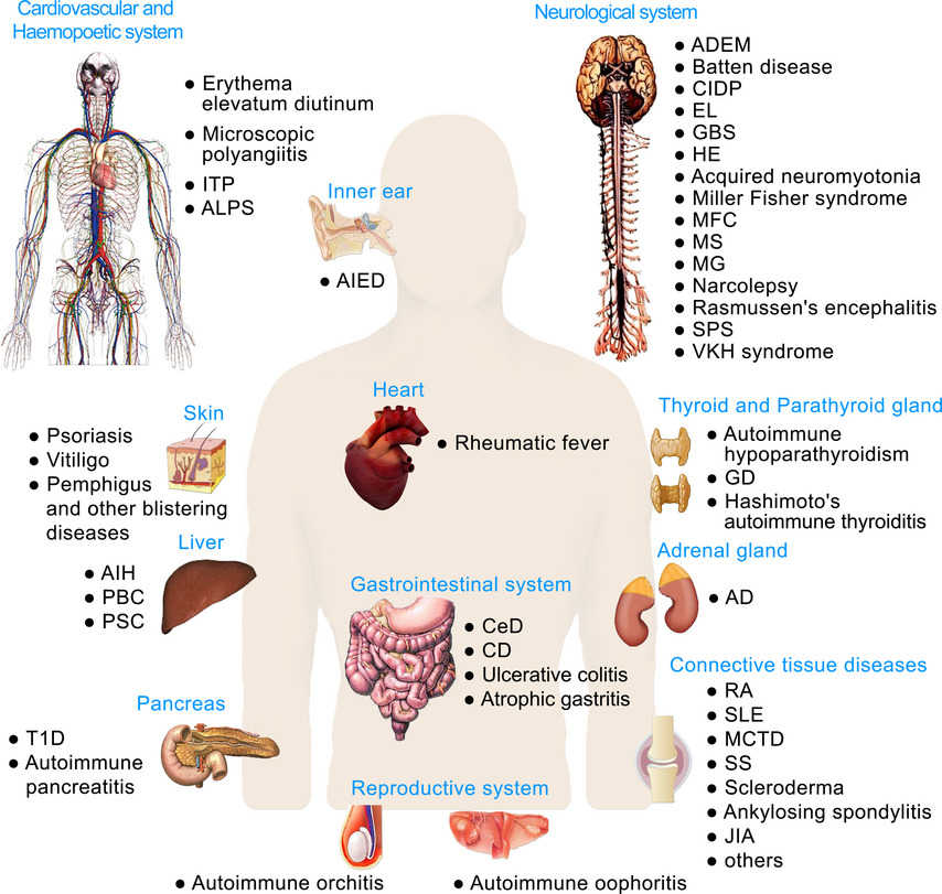
So far, more than 100 different autoimmune diseases have been identified, such as type 1 diabetes, lupus, autoimmune thyroid disease, and rheumatoid arthritis, which together affect over 50 million people in the U.S. Autoimmune diseases are characterized by immune disturbances that cause aberrant B cell and T cell reactivity to normal constituents of the host. Among these diseases, the most prominent immunological manifestation is the production of autoantibodies, which provide valuable biomarkers for diagnosis, classification and disease activity.
 Figure 1: Autoimmune diseases
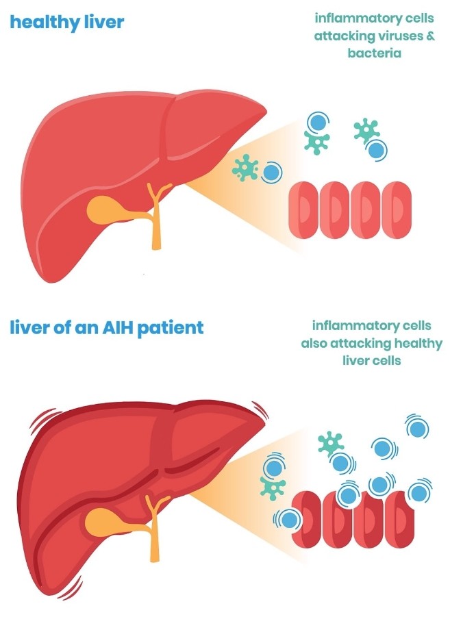
Autoimmune disease happens when the body's natural defense system can't tell the difference between your own cells and foreign cells, causing the body to mistakenly attack normal cells. In general, autoimmune disease results from an interplay between a genetic predisposition and environmental factors.
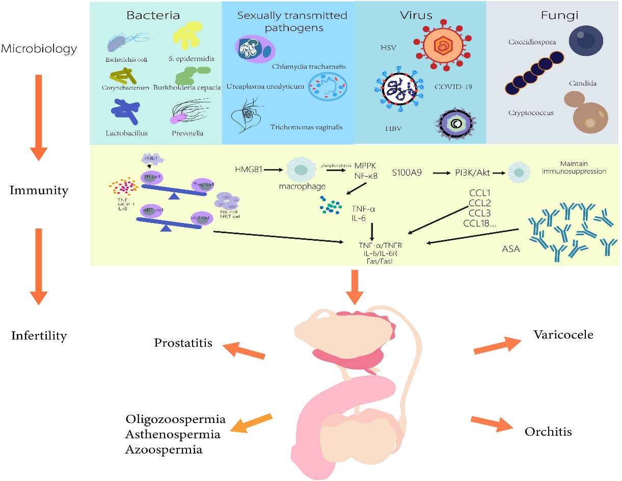
Common environmental factors that may induce autoimmune disease include exogenous compounds (such as cigarette smoke, hormones, vaccines, implants and heavy metals), exogenous microorganisms (viruses) and ultraviolet rays. Exogenous compounds, microorganisms and apoptosis produce autoantigens, which activate B and T cells under the action of antigen-presenting cells and epitope diffusion mechanisms. In addition, the microbiome in the skin, respiratory tract and digestive tract will also stimulate the activation of B and T lymphocytes, eventually leading to the onset of autoimmune diseases.
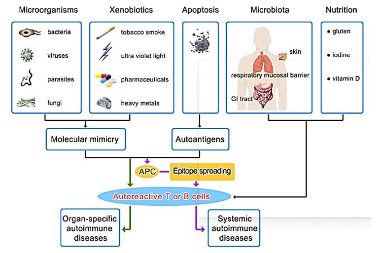 Figure 2: Environmental factors in autoimmunity
Epigenetic inheritance, single gene inheritance and polygene inheritance are three types of inheritance that induce autoimmune disease. The CD40L promoter was hypermethylated in patients with primary biliary cirrhosis. People with T1D have higher levels of insulin-related gene methylation than healthy people. It can be seen that epigenetic modification is closely related to the occurrence of autoimmune diseases. Single-gene autoimmune diseases are rare. In most cases, many autoimmune diseases are caused by the interaction of multiple genes. Moreover, many autoimmune diseases occur with a higher frequency in women than men, with sex representing a major genetic risk factor, for example, SLE, characteristically affects women in their childbearing years.
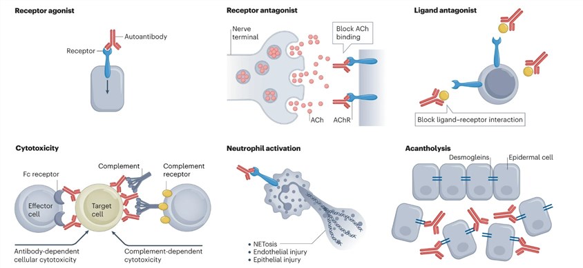 Figure 3: The actions of autoantibodies in immunopathology.
Autoantibodies are widely used in the diagnosis and prognosis of autoimmune diseases. For example, in type 1 diabetes, the most significant autoantibodies are directed to islet cell cytoplasmic (ICA), insulin (IAA), glutamic acid decarboxylase (GADA), and IA2/ICA512 autoantigen (IA2A), have been used to assist in classification and diagnosis. In rheumatic diseases, such as rheumatoid factor (RF) and anti-nuclear antibodies (ANAs) are frequently found in rheumatoid arthritis (< 75%) and systemic lupus erythematosus (SLE) (90–100%), respectively. Graves' disease and Hashimoto's thyroiditis can be easily diagnosed and monitored by anti-thyroid autoantibody levels. Celiac disease can mainly be diagnosed and monitored by anti-tTG and DGP autoantibody tests. Moreover, some autoantibodies may be detected many years before the onset of autoimmune disease. It was reported that over 88% of SLE patients showed at least one SLE autoantibody-positive test (e.g. aANA, antiphospholipid, anti-Ro, anti-La, anti-Sm, anti-nuclear ribonucleoprotein and anti-double-stranded DNA autoantibodies) before the diagnosis of SLE (up to 9.4 years earlier; mean, 3.3 years).
Multiple methods are used in the laboratory to detect the presence of autoantibodies, with descriptive and semi-quantitative or quantitative results.
1. Descriptive and semi-quantitative assays
2. Quantitative assays
3. Biopsy testing in autoimmunity
In SLE, immunofluorescent microscopy of kidney biopsy samples can show the so-called full-house pattern with the presence of immunoglobulins IgG, IgA and IgM and complement in the glomerulus; electron microscopy can further localize immune complexes to either the sub-epithelial or sub-endothelial side of the basement membrane. In contrast to the 'lumpy bumpy' pattern of immunoreactants in lupus nephritis, the immunohistochemistry of kidney biopsy samples from patients with Goodpasture syndrome (also known as anti-GBM disease; a pulmonary-renal syndrome) shows a linear pattern of staining.
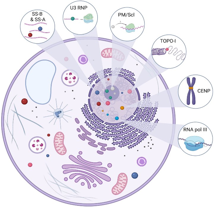
The Antinuclear Antibodies (ANAs) bind to cellular components in the nucleus, including proteins, DNA, RNA, and nucleic acid-protein complexes, and have been the foundation of diagnosis for autoimmune connective tissue disease, including systemic lupus erythematosus (SLE), Sjogren's syndrome, and polymyositis/dermatomyositis.
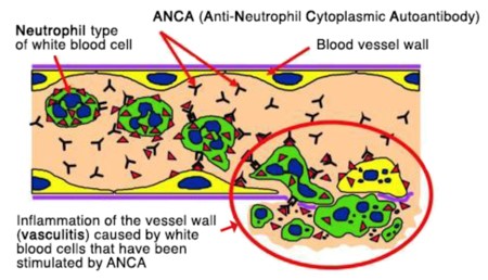
ANCAs attack healthy white blood cells called neutrophils, which can lead to autoimmune vasculitis. There are two main kinds of ANCA: pANCA and cANCA. There are several types of autoimmune vasculitis, such as Granulomatosis with polyangiitis (GPA), Microscopic polyangiitis (MPA), and Eosinophilic granulomatosis with polyangiitis (EGPA), and they all cause inflammation and swelling in your blood vessels. Testing for pANCA can also help diagnose certain types of inflammation.
Type 1 diabetes results from the immune-mediated destruction of insulin-producing pancreatic beta cells, leading to high blood sugar levels that currently require lifelong insulin treatment to keep patients alive. Type 1 diabetes (T1D) Islet autoantibodies that recognize insulin (IAA), glutamic acid decarboxylase (GADA), protein phosphatase-like IA-2 (IA-2A), zinc transporter 8 (ZnT8A), and islet cell cytoplasmic antigen (ICA), are well-validated predictors of risk and disease progression and have been proposed as diagnostic markers of presymptomatic stages of T1D.
In rheumatoid arthritis (RA) the immune system attacks the joints resulting in their damage and destruction and, eventually, disability occurs. Approximately 0.5-1% of adults worldwide suffer from RA. Important autoantibody biomarkers for RA include rheumatoid factor (autoantibody against the fragment-crystallizable (Fc) region of IgG), antibodies against post-translationally modified proteins involving citrullination (ACPA), and carbamylation (anti-CarP antibodies). Immune complexes in the joint may be formed by these autoantibodies, which cause swelling, redness, stiffness and warmth.
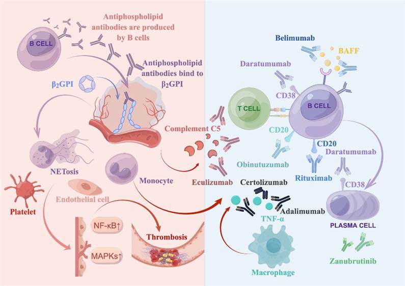
In antiphospholipid syndrome (APS), the immune system produces antiphospholipid antibodies attached to phospholipids, which makes the blood more likely to clot. The blood tests focus on three APS antibodies: anticardiolipin, beta-2 glycoprotein I (β2GPI), and lupus anticoagulant. Lupus anticoagulant test is the strongest predictor for adverse pregnancy-related events. It is more specific than anticardiolipin antibodies in predicting thrombosis. In anticardiolipin antibodies and anti-beta-2-glycoprotein I antibodies IgG and IgM isotypes tests, IgG antibodies correlate better with clinical manifestations than IgM or IgA.
Gangliosides are essential for proper cell signaling, transduction and influences neuroplasticity. Antibodies targeting gangliosides are usually developed as a consequence of molecular mimicry following infections. Binding of antibodies to gangliosides on axonal membranes, nodes of Ranvier, myelin sheath components, Schwann cells, neuromuscular junctions or other neural cell surfaces may elicit inflammatory damage through complement-dependent and independent mechanisms, resulting in nerve conduction blocks and subsequent axonal degeneration. Antiganglioside antibodies are implicated in many neurological disorders including Guillain-Barré syndrome, chronic inflammatory demyelinating polyradiculoneuropathy, multifocal motor neuropathy, and neurodegenerative disorders.
Autoimmune thyroid diseases (AITD) are the most prevalent organ-specific autoimmune diseases (ADs) including Graves' Disease (GD) and Hashimoto Thyroiditis (HT), among others. HT and GD are the major causes of hypothyroidism and hyperthyroidism, respectively. The TPOAb and TgAb are the most common thyroid autoantibodies present in patients with AITD and are associated with complement - mediated cytotoxicity against thyrocytes. There are other less common autoantibodies such as those against the sodium/iodine symporter (NIS) and pendrin, but their clinical utility is limited.
Anti-synthetase syndrome is characterized by autoantibodies against one of many aminoacyl transfer RNA (tRNA) synthetases with clinical features that may include interstitial lung disease (ILD), non-erosive arthritis, myositis, Raynaud's phenomenon, unexplained fever and/or mechanic's hands. The hallmark of anti-synthetase syndrome is the presence of myositis-specific anti-synthetase antibodies, of which anti-Jo-1, an anti-histidyl-tRNA synthetase, is most commonly identified. Less common anti-synthetase antibodies include anti-threonyl (anti-PL7), anti-alanyl (anti-PL12), anti-isoleucyl (anti-OJ), and anti-gylcyl (anti-EJ).
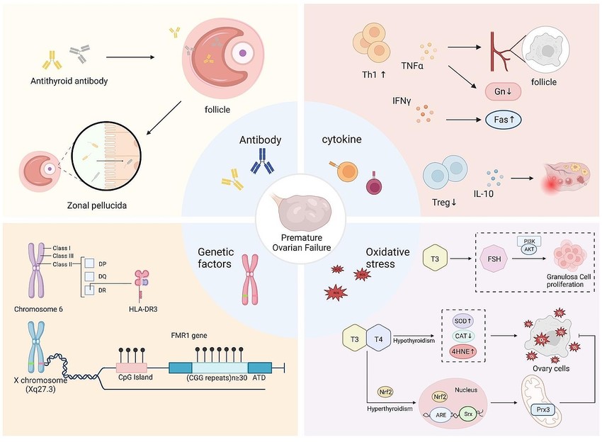
Women with autoimmune diseases have an increased risk of premature ovarian failure. In addition, the autoantibodies may play an important role in fertilization and embryo implantation rates as well as placental development. Clinical symptoms such as repeated miscarriages and increased risk of thrombotic complications during pregnancy are associated with the presence of antiphospholipid antibodies in blood. It increases the risk of pathologies such as pre-eclampsia, RIC and preterm birth. Some studies have seen a lower ovarian reserve in women with uncontrolled MS. Endometriosis has also been found in some autoimmune diseases.
Loading ......
