
Inquiry Basket
 Loading ......
Autoimmune hepatitis (AIH) is a liver disorder characterized by persistent inflammation, hepatocellular damage, and liver fibrosis, due to an abnormal immune response. It is classified into two main types based on serological markers and histological features: AIH type 1 (AIH-1) and AIH type 2 (AIH-2). AIH is more common in women. The clinical presentation ranges from asymptomatic liver enzyme elevation to severe acute liver failure.
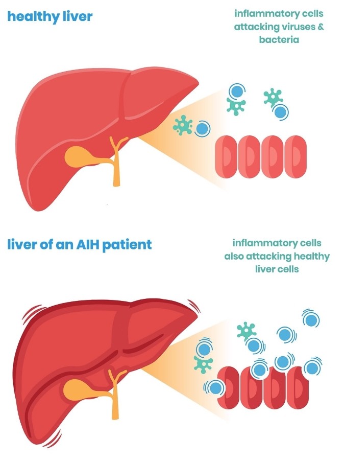 Fig. 1 How autoimmune hepatitis affects the liver
The etiology of AIH is multifactorial, involving genetic predisposition, environmental triggers, and dysregulated immune responses. AIH-1 is commonly associated with HLA-DR3 and HLA-DR4, while AIH-2 has been linked with HLA-DR8. These alleles are involved in antigen presentation and may influence the immune system's ability to recognize and respond to liver antigens. Specific single nucleotide polymorphisms (SNPs) have been associated with an increased risk of AIH. For example, variations in the genes encoding interleukin-10 (IL-10) and tumor necrosis factor receptor (TNFR) have been linked to AIH susceptibility. Environmental factors can act as triggers in genetically predisposed individuals, including viral Infections (Hepatitis A, hepatitis B, and Epstein-Barr virus (EBV) infections have been implicated). Minocycline and isoniazid have been associated with drug-induced autoimmune hepatitis. Estrogen and other sex hormones might influence immune responses and contribute to the higher prevalence of AIH in females.
The dysregulation of immune responses is central to the pathogenesis of AIH. Several autoantigens have been identified in AIH, and their presentation to the immune system is crucial for the disease development of AIH. These include:
In addition to autoantigens, pro-inflammatory cytokines such as tumor necrosis factor-alpha (TNF-α), interleukin-6 (IL-6), and interleukin-17 (IL-17) are involved in liver inflammation and fibrosis. The cytokine milieu influences the progression and severity of liver damage.
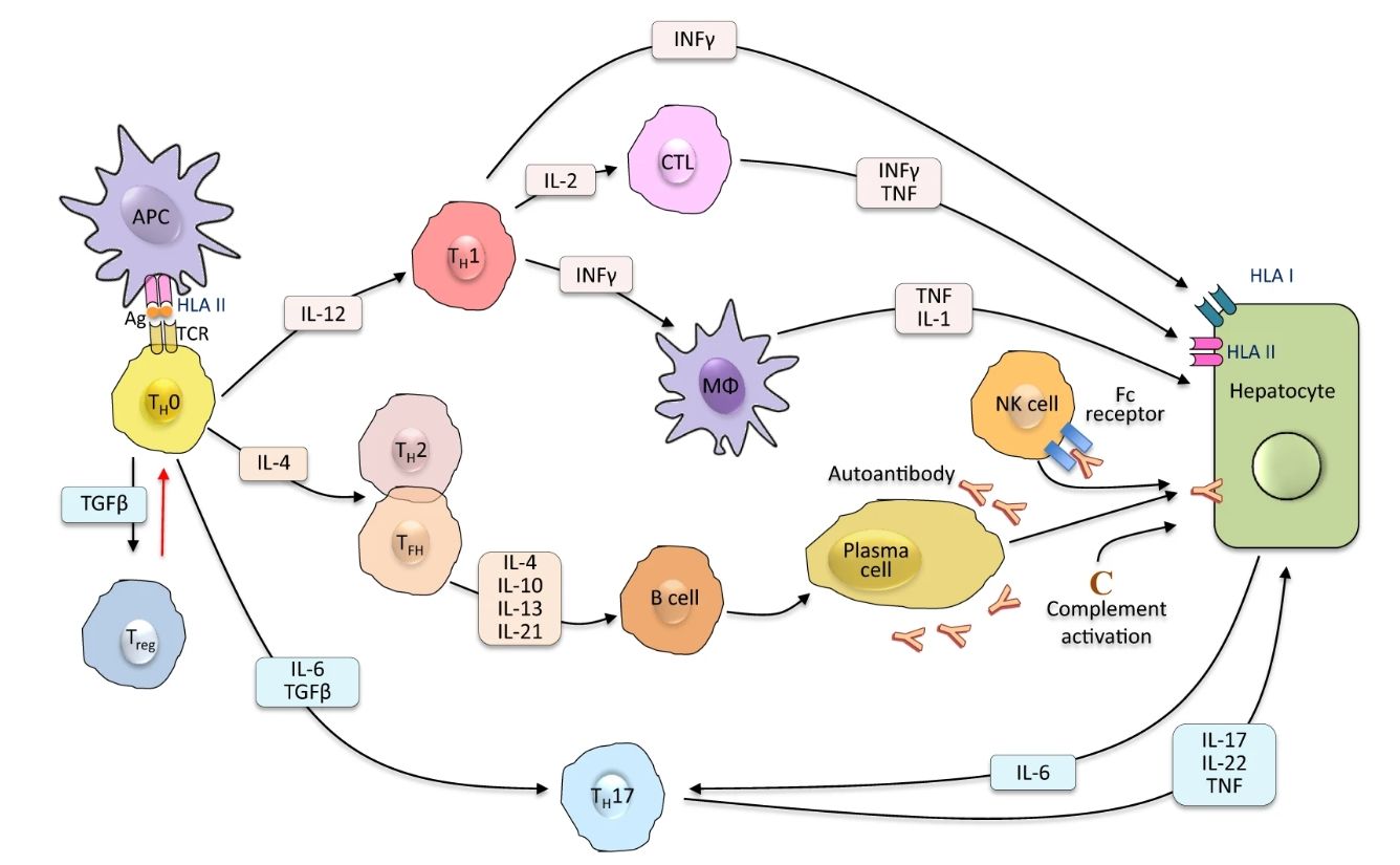 Fig. 2 Autoimmune attack to the liver cell
AIH is diagnosed based on the presence of hypergammaglobulinemia, serum autoantibodies and histological features of interface hepatitis. Key markers include:
Table 1: Autoantibodies in different two AIHs
| Anti-LKM-1 Antibodies | |||
| CABT-L2414 | Humanized Anti-Human LKM-1 Monoclonal antibody, clone H8B4 | IEP, WB | Inquiry |
| Anti-LC1 Antibodies | |||
| CABT-L2416 | Humanized Anti-Human LC1 Monoclonal antibdoy, clone D8F5 | IEP, WB | Inquiry |
| Anti-SEPSECS Antibodies | |||
| CABT-Z519H | Human Anti-Human SLA/LP Chimeric Monoclonal antibody, clone 47311 | ELISA | Inquiry |
| DPABH-04282 | Rabbit Anti-Human SEPSECS Polyclonal antibody | WB, IP, ELISA | Inquiry |
| DPABH-10995 | Rabbit Anti-Human SEPSECS (aa 443-474) Polyclonal antibody | WB | Inquiry |
| Anti-SMA Antibodies | |||
| CABT-L1593 | Rabbit Anti-Human SMA Monoclonal antibody, clone 28I20M46 | ICC, IHC-P, IF, WB | Inquiry |
| Anti-Nuclear Antigen Antibodies | |||
| DCABY-098 | Mouse Anti-Nuclear antigen Monoclonal antibody, clone 69-26 | IF, IHC-P | Inquiry |
| Anti-gp210 Antibodies | |||
| CABT-BL1650 | Mouse Anti-gp210 Monoclonal antibody, clone AGP26.10 | WB | Inquiry |
| Anti-SP100 Antibodies | |||
| CABT-L2418 | Humanized Anti-Human SP100 Monoclonal antibody, clone C7D6 | IEP, WB | Inquiry |
| CABT-B1289 | Mouse Anti-Human SP100 (N-terminal half) Monoclonal antibody, clone 3I2.2 | WB, ICC | Inquiry |
| DPABH-06742 | Mouse Anti-Human SP100 (full length) Polyclonal antibody | WB, IHC-P | Inquiry |
| CABT-L2093 | Rabbit Anti-Human Sp100 Polyclonal antibody | WB | Inquiry |
| CPBT-47125RH | Rabbit Anti-Human SP100 (aa 250-350) Polyclonal antibody | IHC-P, WB | Inquiry |
| DPABT-H21500H | Rabbit Anti-Human SP100 (aa 209-227) Polyclonal antibody | IF, WB | Inquiry |
Loading ......
