
Inquiry Basket
 Loading ......
The Antinuclear Antibodies (ANAs) are a class of antibodies that bind to cellular components in the nucleus, including proteins, DNA, RNA, and nucleic acid-protein complexes and have been the foundation of diagnosis for autoimmune connective tissue disease, including systemic lupus erythematosus (SLE), Sjogren's syndrome, and polymyositis/dermatomyositis. A certain percentage of the healthy population has detectable levels of ANAs, while increased titers are characteristic of individuals with connective tissue disorders. Thus, the sensitivity and specificity of the methods used to detect ANAs are crucial to diagnosis.
Anti-dsDNA antibody is a representative ANA, autoantibodies against other targets include Sm, dsDNA, nucleosomes, histones, P0, PCNA, SSA, Ro52, SSB/La, Scl-70, Jo-1, PM/Scl, CENP-B, nRNP/Sm, AMA-M2, etc.
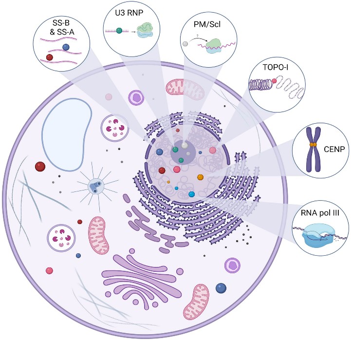 Figure 1: Main antigenic targets of ANAs
The antinuclear antibodies may bind to double-stranded DNA (anti-dsDNA), and studies suggest the antibodies are formed during the incomplete removal of cellular material during apoptosis. Additionally, anti-Sm antibodies bind to the Smith protein, a protein contained within small nuclear ribonucleoprotein (snRNP) particles. Scl-70 antibodies interfere with DNA replication by binding to Topoisomerase I, and anti-centromere antibodies affect cell division by binding to centromeres during interphase. Antibodies to Jo-1 prevent histidine binding to tRNA during protein synthesis by targeting histidyl-tRNA synthetase. Additionally, antibodies may target the Ro/SSA antigen, an amino acid sequence that binds to double-stranded and single-stranded DNA. The suspected mechanism is that they may bind to viral DNA of the Epstein Barr virus (EBV) with molecular mimicry, later causing autoimmune disease. Unlike Ro, La/SSB is a protein found primarily in the nucleus. But similar to Ro, La is also known to bind to nuclear material from EBV.
ANA-IIF is the current most endorsed technique for ANA detection. HEp-2 cells are commonly used as a substrate and can present distinct patterns of fluorescence depending on the antibody in the human serum and the localization of the antigen in the cell, including homogeneous, speckled, nucleolar, nuclear membranous, centromeric, nuclear dot, and pleomorphic. In addition, the level of autoantibodies is reported as a titer. Positive autoantibody titers at a dilution equal to or greater than 1:160 are usually considered clinically significant. Moreover, enzyme-linked immunosorbent assays (ELISA) method offers confirmatory testing for specific ANAs and quantitative screening for specific ANAs.There are two types of EIA or ELISA methods currently used for ANA testing. One is called generic assay which detects ANA of broad specificity similar to IF-ANA and other is antigen specific assay that detects ANA and reacts with a single autoantigen i.e. dsDNA, SS-A/Ro, SS-B/La, Scl-70, Sm, Sm/RNP etc. The usage of a combination of IIF and ELISA shows more clinical value.
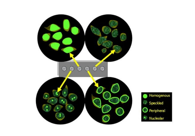 Figure 2: Diagrammatic representation of common nuclear patterns observed under fluorescence microscopy.
| ANA pattern | Antigen | Associated diseases |
| Speckled | ENA, RNP, Sm, Ro/SS-A, SSB, Scl-70, Jo-1, Ribosomal P | SLE, Mixed CTD, SS, Primary Sjogren's syndrome, PM |
| Homogenous | dsDNA, Histone | SLE, Drug induced SLE |
| Peripheral (rim) | RNP, Sm, Ro/SS-A | SLE, SS |
| Nucleolar | PM/Scl100, Scl-70, FBL | SS, PM |
| Centromere | CENP-B, | Limited SS |
Table 1. Common IF-ANA patterns associated with specific diseases
Loading ......
