
Inquiry Basket
 Loading ......
Antineutrophil cytoplasmic antibodies (ANCA) are autoantibodies directed against antigens found in the cytoplasmic granules of neutrophils and monocytes. There are two main kinds of ANCA: pANCA and cANCA. The c-ANCA antigen is maily (over 90%) proteinase 3 (PR3), other antigens exist for c-ANCA (atypical). p-ANCA antigens include myeloperoxidase (MPO) and bacterial permeability increasing factor Bactericidal/permeability-increasing protein (BPI). ANCA testing is usually performed to help to diagnose and monitor inflammatory activity in Wegener granulomatosis, microscopic polyangiitis and its renal-limited variant (pauci-immune crescentic glomerulonephritis), and Churg-Strauss syndrome. Testing for pANCA can also help diagnose certain types of inflammatory bowel disease (IBD).
Antineutrophil cytoplasmic antibodies (ANCA) are a sensitive and specific marker for ANCA-associated systemic vasculitis, including granulomatosis with polyangiitis, microscopic polyangiitis, primary pauci-immune necrotizing crescentic glomerulonephritis (a type of renal-limited microscopic polyangiitis), eosinophilic granulomatosis with polyangiitis and drug induced vasculitides. PR3 directed c-ANCA is present in 80-90% of granulomatosis with polyangiitis, 20-40% of microscopic polyangiitis, 20-40% of pauci-immune crescentic glomerulonephritis and 35% of eosinophilic granulomatosis with polyangiitis. c-ANCA (atypical) is present in 80% of cystic fibrosis (with BPI as the target antigen) and also in inflammatory bowel disease, primary sclerosing cholangitis and rheumatoid arthritis. p-ANCA with MPO specificity is found in 50% of microscopic polyangiitis, 50% of primary pauci-immune necrotizing crescentic glomerulonephritis and 35% of eosinophilic granulomatosis with polyangiitis. In addition, the ANCA‐positive rate is much higher in patients with type 1 diabetes mellitus than in healthy individuals. Levamisole can cause an ANCA positive vasculitis (1).
ANCA are not only a measureable blood marker in patients with ANCA-associated vasculitis but they are harmful autoantibodies that are directly involved in small blood vessel damage. MPO and PR3 specific ANCA can activate neutrophils and monocytes through their Fc and Fab'2 receptors, which can be enhanced by cytokines that cause neutrophils to display MPO and PR3 on their surface. The activated neutrophils can then adhere to endothelial cells, resulting in damage to the endothelium via the induction of necrosis and apoptosis. Furthermore, neutrophils release chemoattractive signaling molecules that recruit more neutrophils to the endothelium, acting as a positive feedback loop. Animal models have shown that MPO antibodies can induce necrotizing crescentic glomerulonephritis and systemic small vessel vasculitis. ANCAs induce excess activation of neutrophils, resulting in the production of neutrophil extracellular traps (NETs), which cause damage to small blood vessels. In addition, ANCA reactive B-cells can be found in circulation in patients with AAV, an alternative hypothesis has been proposed assigning a direct pathogenic role of these cells, whereby activated neutrophils and ANCA-reactive B-cells engage in intercellular cross-talk, which leads not only to neutrophil degranulation and inflammation but also to the proliferation and differentiation of ANCA-reactive B-cells (2).
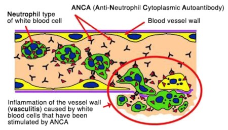 Fig. 1 Diagram showing ANCA-induced 'activation' of neutrophils causing inflammation of the blood vessel wall
A conventional screening test to detect ANCA in the serum is indirect immunofluorescence study, and subsequently confirmed by enzyme-linked immunosorbent assay. A positive staining of ANCA can be classified into three main categories based on the staining patterns: cytoplasmic, perinuclear, and atypical. Patients with granulomatosis with polyangiitis (GPA) mostly have a positive cytoplasmic staining pattern (c-ANCA) whilst a perinuclear pattern (p-ANCA) is more common in microscopic polyangiitis (MPA) and eosinophilic granulomatosis with polyangiitis (EGPA) patients. Atypical pattern (a-ANCA) is rarely seen in patients with systemic small-vessel vasculitis but it can be found in other conditions.
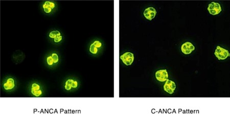 Figure 2. Indirect immunofluorescence of ANCA
Systemic vasculitis with ANCA [ANCA-associated vasculitides (AAV)] is a subgroup of life-threatening inflammatory disorders affecting small- to medium-sized vessels. Immunosuppressants and glucocorticoids represent the current therapeutic mainstay, but with a 25% present with severe adverse events, and standard therapy does not sustain remission. It has turned ANCA-associated vasculitis (AAV) into chronic, relapsing disorders. Therefore, an unmet need for safer and more effective therapies has prompted interest in biological agents.
A reasonable strategy to suppress the autoimmune cellular response might be the blockage of costimulating molecules that intervene in the antigen presenting cell-dependent T cell activation which is thought to be the initial step in Wegener granulomatosis granuloma formation. At the vasculitic pole of the spectrum, monoclonal antibodies and fusion proteins aimed at inhibiting ANCA, suppressing neutrophil priming and endothelial activation, and interfering in the neutrophil-endothelium adhesion cascade or neutrophil degranulation might also prove fundamental in healing vasculitic damage.
References
| Cat. No. | Product name | Application | |
| CABT-L6305 | Human Anti-Human MPO monoclonal antibody, clone D29 | ELISA | Inquiry |
| CABT-Z262R | Recombinant Rabbit Anti-Human MPO Monoclonal Antibody, clone CQ7215 | IHC | Inquiry |
| DCABH-2947 | Rabbit Anti-Human MPO monoclonal antibody, clone KN21-69 | WB, ICC/IF, IHC | Inquiry |
| DCABH-5222 | Mouse Anti-Mouse/Rat MPO monoclonal antibody, clone 9G5 | IHC, FC | Inquiry |
| DCAB-TJ224 | Mouse Anti-Human MPO monoclonal antibody, clone 29C8 | ELISA(Cap) | Inquiry |
| DCAB-TJ225 | Mouse Anti-Human MPO monoclonal antibody, clone 27F4 | ELISA(Det) | Inquiry |
| DMAB-JXL2347 | Mouse Anti-Human MPO Monoclonal Antibody, clone 833 | ELISA | Inquiry |
| CABT-L0219Y | Human Anti-Human MPO polyclonal antibody | ELISA, IF | Inquiry |
| DPABH-05549 | Rabbit Anti-Human MPO Polyclonal Antibody | WB, IHC | Inquiry |
| DPAB-JXL23178 | Rabbit Anti-Human MPO polyclonal antibody | ELISA, WB | Inquiry |
| Cat. No. | Product name | Nature | Application | |
| DAG-WT2500 | Myeloperoxidase (MPO) control | Synthetic | IA | Inquiry |
| DAG-WT3245 | Recombinant Human MPO [His] | Recombinant Expression system: CHO cells | ELISA | Inquiry |
| DAG-WT1043 | Recombinant Myeloperoxidase [His] | Recombinant Expression system: HEK293 cells | Control | Inquiry |
| DAG627 | Human Myeloperoxidase | Native | WB, ELISA | Inquiry |
| DAGA-868 | Myeloperoxidase (pANCA) (>95%) | Native | ELISA | Inquiry |
| DAGA-874 | Myeloperoxidase antigen isoform A (>96%) | Native | Controls, Calibrators, ELISA, Blotting | Inquiry |
| DAGA-875 | Myeloperoxidase antigen isoform B (>96%) | Native | Controls, Calibrators, ELISA, Blotting | Inquiry |
| DAGA-876 | Myeloperoxidase antigen isoform C (>96%) | Native | Controls, Calibrators, ELISA, Blotting | Inquiry |
| Cat. No. | Product name | Detection Sample | Application | |
| DEIA1820 | Human MPO Antibody ELISA Kit | Serum, plasma | Quantitative | Inquiry |
| DEIA5691 | Mouse MPO ELISA kit | Plasma, and cell culture supernatants | Quantitative | Inquiry |
| DEIA-XY2177 | Rat MPO ELISA kit | Cell culture medium, plasma, tissue homogenates | Quantitative | Inquiry |
| DEIA1698 | Human Myeloperoxidase IgG ELISA Kit | Serum | Semi-quantitative, Qualitative | Inquiry |
| DEIA2776 | Human Myeloperoxidase ELISA Kit | Cell culture supernatants, cell lysates, serum, platelet-poor plasma, saliva, urine | Quantitative | Inquiry |
| DEIA6443 | Human Myeloperoxidase ELISA Kit | Biological fluids | Quantitative | Inquiry |
| Cat. No. | Product name | Detection Sample | |
| DTS633 | Myeloperoxidase Rapid Test | Whole blood | Inquiry |
| Cat. No. | Product Name | Application | |
| CABT-L0218Y | Human Anti-Human protienase-3/PR3 polyclonal antibody | ELISA | Inquiry |
| CABT-L6003 | Human Anti Human proteinase-3 monoclonal antibody, clone D21 | ELISA | Inquiry |
| CABT-B10680 | Mouse anti-Human MPND monoclonal antibody, clone 2D23 | WB, sELISA, ELISA | Inquiry |
| CABT-B1482 | Mouse Anti-Human PRTN monoclonal antibody, clone NDQS4-4 | WB, FC, ELISA | Inquiry |
| CABT-B1483 | Mouse Anti-Human PRTN monoclonal antibody, clone NDQS4-8 | WB, Inhib, FuncS, IP, FC | Inquiry |
| DCABH-10226 | Mouse Anti-Human PR3 monoclonal antibody, clone XHN3 [FITC] | WB, IHC, FC, ELISA | Inquiry |
| DCABH-16540 | Mouse Anti-Human PR3 monoclonal antibody, clone 4C5 | WB, ELISA | Inquiry |
| DCABH-2820 | Rabbit Anti-Human PR3 monoclonal antibody, clone FQS7388 | WB, IHC-P | Inquiry |
| Cat. No. | Product name | Nature | Application | |
| DAG628 | Human Proteinase 3 | Native | ELISA | Inquiry |
| DAG-WT2251 | Recombinant Human Proteinase 3 [His] | Recombinant Expression System: Mammalian cells | IA | Inquiry |
| DAG-WT2781 | Recombinant Human Proteinase 3 [His] | Recombinant Expression System: HEK293 cells | ELISA | Inquiry |
| Cat. No. | Product name | Detection Sample | Application | |
| DEIA3141 | Human ANCA-C (PR3) ELISA Kit | Serum, plasma | Quantitative | Inquiry |
| DEIA05726 | Human Anti-PR-3 (C-ANCA) IgG ELISA Kit | Serum | Semi-quantitative, Qualitative | Inquiry |
| DEIA1700 | Human Proteinase-3 IgG ELISA Kit | Serum | Semi-quantitative, Qualitative | Inquiry |
| DEIA-BJ2253 | Rat Proteinase 3 antibody ELISA Kit | Serum, plasma, cell culture supernatants, body fluid and tissue homogenate | Quantitative | Inquiry |
Loading ......
