
Inquiry Basket
 Loading ......
Myasthenia gravis (MG) is a prototypical antibody-mediated autoimmune disease affecting the neuromuscular junction (NMJ). In 2021, the diagnosed prevalence and incidence of MG in the United States were calculated to be 37.0 per 100,000 persons and 3.1 per 100,000 persons, respectively. Patients with MG exhibit a spectrum of weakness, ranging from limited ocular muscle involvement to life-threatening respiratory failure. The disease is characterized by the presence of specific antibodies that target components of the NMJ, primarily the acetylcholine receptor (AChR) and other antigens such as muscle-specific kinase (MuSK) and lipoprotein-related protein 4 (LRP4). Detecting pathogenic antibodies against these synaptic molecules in a patient with the typical clinical features is virtually diagnostic of MG and helps define the disease subtypes.
 Fig. 1 Structure of the NMJ.
(Kaminski HJ.; et al. J Clin Invest. 2024)
MG is a neuromuscular disorder that arises from disrupted communication between nerves and muscles. Typically, acetylcholine binds to receptors in the muscles, initiating contractions. However, in MG, antibodies interfere with, modify, or destroy these receptors, hindering appropriate muscle activation. Although most instances involve antibodies against acetylcholine receptors, some patients have antibodies against MuSK or LRP4, which also disturb neuromuscular transmission. The thymus gland, which is crucial for immune system maturation, may play a role in MG development. In many adults with the condition, it is also accompanied by the abnormal enlargement of the thymus or thymoma. Scientists believe the thymus may send incorrect signals to immune cells, leading to the production of harmful antibodies. Additionally, certain medications, including statins used to lower cholesterol, may worsen MG symptoms.
 Fig. 2 Pathogenic mechanisms of MG autoantibodies at the NMJ.
(Koneczny I & Herbst R. Cells. 2019)
Approximately 85% of MG patients have autoantibodies against AChRs, primarily of the IgG1 and IgG3 subclasses targeting the main immunogenic region (MIR). These antibodies induce pathogenicity through three main mechanisms:
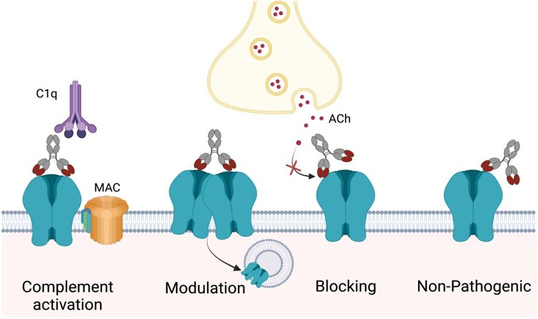 Fig. 3 Mechanisms of AChR autoantibody-mediated pathology.
(Masi G & O'Connor KC. Curr Opin Neurol. 2022)
Activation of the Complement Cascade: Antibodies bind to AChR, activating the classical complement pathway via C1q, leading to (membrane attack complex) MAC formation and postsynaptic membrane lysis, causing severe NMJ damage.
Antibody-Induced Endocytosis of AChRs: Antibodies cross-link AChRs, promoting their internalization and degradation, which reduces AChR density at the NMJ.
Direct Functional Blockade of AChRs: Antibodies directly inhibit AChR function by preventing acetylcholine binding or blocking the ion channel.
MuSK antibodies in myasthenia gravis (MG) differ fundamentally from acetylcholine receptor (AChR) antibodies. They are predominantly of the IgG4 subclass, which does not activate the classical complement pathway or immune cells due to structural differences in the Fc region. MuSK antibodies primarily bind to the first Ig-like domain of MuSK, particularly around Ile96, disrupting the interaction between MuSK and Lrp4. This blockade reduces MuSK phosphorylation and interrupts the Agrin-Lrp4-MuSK-Dok-7 signaling axis, leading to decreased densities of acetylcholine receptors (AChRs) at the synapse and impaired neuromuscular transmission.
LRP4 functions as a critical protein at the NMJ by binding agrin and initiating AChR clustering with the assistance of MuSK. The LRP4 antibodies are primarily IgG1 and IgG2 subtypes and follow similar clinical presentation to a mild form of early-onset MG, inhibit clustering of AChR, and appear not to strongly activate complement. Additionally, Some studies on Agrin autoantibodies suggest they may block Lrp4-Agrin interaction, impairing MuSK activation.
 Fig. 4 Structure of Agrin, Lrp4, and MuSK.
(Koneczny I & Herbst R. Cells. 2019)
Detecting established pathogenic autoantibodies against synaptic molecules in patients with typical clinical features can diagnose MG and helps define disease subtypes. About 80-90% of MG patients have detectable serum antibodies against AChRs with 40-70% of the remaining patients being positive for anti-MuSK antibodies and 2-50% for anti-LRP4 antibodies. Besides the above, agrin, collagen Q, and voltage-gated potassium channel Kv1.4 are also can be detected. At present, commercial assays are available for detecting AChR-Ab, MuSK-Ab, and LRP4-Ab, with ongoing refinements, while other techniques remain limited to leading research institutions. Although not directly pathogenic, several other autoantibodies are useful in assessing thymoma-associated MG and late-onset MG. Several commonly used detection methods and autoantibody prevalence are summarized as follow.
| Autoantigen | Detection method | % of MG patients | % of dSN-MG patients |
| AChR | RIPA | 80–85% | N.A. |
| Clustered AChR | CBA | N.T. | ~20% (4–60%) |
| MuSK | RIPA | ~6% (2–3% in Japanese) | N.A. |
| MuSK | CBA | N.T. | 13% |
| LRP4 | CBA | ~2% | ~19% |
| Titin | ELISA | 20–30% (90% in thymoma EOMG) | 0–3% |
| Titin | RIPA | ~41% | 13.40% |
| RyR | ELISA | ~ 14% in LOMG (75% in thymoma MG) | N.T. |
| Agrin | ELISA/CBA | 2–15% | 0–50% |
| Kv1.4 | IP and SDS-PAGE | 10–20% | 0 |
| Rapsyn | Immunoblots | 11% | 17% |
| Cortactin | ELISA, WB | 5–10% | ~20% |
| ColQ | CBA | 3% | 3.40% |
References
| AChR Antibodies | ||||
| Target | Cat. No. | Product Name | Application | |
| AChR | CABT-L4419 | Rat Anti-Human AChR Monoclonal antibody, clone Mab35 (TIB-175) | IF | Inquiry |
| DMAB-JXL2334 | Rat Anti-Human AChR Monoclonal antibody, clone Mab192 | BL, WB, ELISA | Inquiry | |
| DMAB-JXL2335 | Rat Anti-Human AChR Monoclonal antibody, clone Mab198 | IA | Inquiry | |
| DMAB-JXL2335H | Humanized Anti-Human AChR monoclonal antibody, clone Mab198 | IA | Inquiry | |
| DMAB-JXL2334H | Humanized Anti-Human AChR monoclonal antibody, clone Mab192 | BL, WB, ELISA | Inquiry | |
| DMAB-JXL2335M | Mouse Anti-Human AChR monoclonal antibody, clone Mab198 | IA | Inquiry | |
| DMAB-JXL2334M | Mouse Anti-Human AChR monoclonal antibody, clone Mab192 | BL, WB, ELISA | Inquiry | |
| CABT-B9172 | Mouse Anti-Rat Acetylcholine Receptor α monoclonal antibody, clone 37 | WB, IF | Inquiry | |
| MuSK Antibodies | ||||
| Target | Cat. No. | Product Name | Application | |
| MuSK | DCABH-12482 | Mouse Anti-Human MuSK monoclonal antibody, clone 20B5 | IHC-P, IF, ELISA | Inquiry |
| DMABC-JX084 | Rabbit Anti-Human MuSK monoclonal antibody, clone 39 | WB, IHC-P | Inquiry | |
| DPABC-JX050 | Rabbit Anti-Human MuSK (Phospho-Tyr755) polyclonal antibody | WB | Inquiry | |
| DPABC-JX051 | Rabbit Anti-Human MuSK (aa 424-783) polyclonal antibody | WB, ELISA | Inquiry | |
| DPABC-JX052 | Rabbit Anti-Human MuSK (aa 231-330) polyclonal antibody | WB, IHC, IF/ICC | Inquiry | |
| DPAB-DC785 | Rabbit Anti-Mouse MuSK (C-terminal) polyclonal antibody | WB, ELISA | Inquiry | |
| LRP4 Antibodies | ||||
| Target | Cat. No. | Product Name | Application | |
| LRP4 | CABT-L0348Y | Mouse Anti-Mouse LRP4 (extracellular) monoclonal antibody, clone O308/38S | WB, ICC, IHC | Inquiry |
| DMABC-JX085 | Mouse Anti-Mouse LRP4 monoclonal antibody, clone 36 | WB | Inquiry | |
| DPABH-21199 | Rabbit Anti-Human LRP4 Polyclonal Antibody | IHC | Inquiry | |
| DPABC-JX053 | Rabbit Anti-Human LRP4 (aa 1500-1725) polyclonal antibody | WB, IHC | Inquiry | |
| DPABC-JX054 | Rabbit Anti-Human LRP4 (aa 1621-1720) polyclonal antibody | ELISA, IF/ICC | Inquiry | |
| DPABH-18692 | Goat Anti-Human LRP4 (aa 1765-1777) polyclonal antibody | WB | Inquiry | |
| Agrin Antibodies | ||||
| Target | Cat. No. | Product Name | Application | |
| Agrin | CPBT-51061RH | Rabbit Anti-Human AGRN Polyclonal Antibody | IHC-P | Inquiry |
| DPABC-JX055 | Rabbit Anti-Human AGRN (aa 1151-1245) polyclonal antibody | IHC, IF | Inquiry | |
| DPABC-JX056 | Rabbit Anti-Human AGRN (aa 30-350) polyclonal antibody | WB, IF/ICC, ELISA | Inquiry | |
| DPABC-JX057 | Rabbit Anti-Human AGRN (aa 968-1130) polyclonal antibody | WB, IHC, IF, ELISA | Inquiry | |
| DPABY-340 | Goat Anti-Rat AGRN polyclonal antibody | WB, IHC, ELISA(Cap), Neut | Inquiry | |
| DPABY-702 | Goat Anti-Rat AGRN polyclonal antibody [Biotin] | WB, ELISA(Det) | Inquiry | |
| DMAB9488MC | Anti-Chicken AGRN Monoclonal antibody, clone 6D2 | WB, AP, IHC | Inquiry | |
| Titin Antibodies | ||||
| Target | Cat. No. | Product Name | Application | |
| Titin | DCABH-9509 | Mouse Anti-TTN monoclonal antibody, clone U22 | WB, ICC/IF, IHC | Inquiry |
| DMABC-JX086 | Mouse Anti-Human TTN monoclonal antibody, clone F5 | WB, IP, IF, ELISA | Inquiry | |
| DPABC-JX058 | Rabbit Anti-Human TTN (aa 2601-2800) polyclonal antibody | IHC, FC, IF | Inquiry | |
| DPABC-JX059 | Rabbit Anti-Human TTN (aa 5347-5604) polyclonal antibody | ELISA, IF/ICC | Inquiry | |
| DPABC-JX060 | Rabbit Anti-Human TTN (aa 5398-5604) polyclonal antibody | ELISA, IHC, IF | Inquiry | |
| DPABC-JX061 | Rabbit Anti-Human TTN (aa 161-210) polyclonal antibody | IHC-P | Inquiry | |
| DPAB-DC3136 | Mouse Anti-Human TTN (aa 1-110) polyclonal antibody | WB, ELISA | Inquiry | |
| RyR Antibodies | ||||
| Target | Cat. No. | Product Name | Application | |
| RyR | DCABY-836 | Mouse Anti-Human RYR1 monoclonal antibody, clone SZS.2 (H-2) | IHC-Fr, ELISA, FC, WB, IF | Inquiry |
| DPABB-JX228 | Rabbit Anti-Human RYR1 (aa 1-534) polyclonal antibody | ELISA, IF | Inquiry | |
| DPABB-JX229 | Rabbit Anti-Human RYR1 (aa 1354-1403) polyclonal antibody | Dot, IHC-P, FC | Inquiry | |
| DPABB-JX230 | Rabbit Anti-Human RYR1 (aa 4701-4800) polyclonal antibody | IHC, IF, ELISA | Inquiry | |
| DPABB-JX231 | Rabbit Anti-Human RYR1 (aa 800-900) polyclonal antibody | WB, ELISA | Inquiry | |
| DPAB-DC2766 | Rabbit Anti-Human RYR1 (C-terminal) polyclonal antibody | WB, ELISA | Inquiry | |
| DPABC-JX062 | Rabbit Anti-Human RYR2 (aa 1471-1520) polyclonal antibody | WB, IF/ICC, IHC-P, FC | Inquiry | |
| DPABC-JX063 | Rabbit Anti-Human RYR2 (aa 901-1192) polyclonal antibody | ELISA, IHC, IF | Inquiry | |
Loading ......
