
Inquiry Basket
 Loading ......
Protein reduction is a fundamental process that plays a pivotal role in various areas of biological research and biotechnology. It involves the reduction of disulfide bonds within proteins, leading to the breakage of these bonds and the conversion of oxidized cysteine residues to their reduced form. Protein reduction is a crucial step in simplifying protein structures and unraveling their functions. By reducing proteins, researchers can obtain valuable insights into their primary structures, interactions, and biochemical properties. This knowledge is instrumental in elucidating disease mechanisms, designing targeted therapeutics, and optimizing protein-based applications.
Learn more about Cysteine Proteases and Regulators
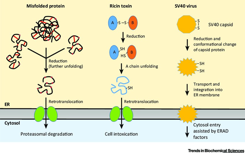 Figure 1. Protein reduction in endoplasmic reticulum (ER)-associated degradation (ERAD) and cytosolic entry of toxins and viruses.
(Source: Ellgaard, L. et al., 2018)
Protein reduction in the endoplasmic reticulum (ER) is a critical process that ensures proper protein folding and quality control in eukaryotic cells. During the synthesis of secretory and membrane proteins, disulfide bonds form within the ER lumen, and the process of protein reduction is crucial for their correct folding and functionality. Here are some key aspects of protein reduction in the ER:
Inside the ER, a distinct oxidative environment exists due to the presence of enzymes, such as protein disulfide isomerases (PDIs), that facilitate protein folding and disulfide bond formation. As nascent proteins enter the ER, cysteine residues are catalyzed to form disulfide bonds, which contribute to the stability and proper folding of secretory and membrane proteins.
PDIs are key enzymes involved in protein reduction and oxidative protein folding within the ER. PDIs catalyze disulfide bond formation, isomerization, and reduction, ensuring the correct pairing of cysteine residues during protein folding. They play a vital role in maintaining the redox balance and quality control of proteins within the ER.
Alongside disulfide bond formation, proper protein folding in the ER is facilitated by a network of molecular chaperones. Chaperones assist in the folding, assembly, and quality control of newly synthesized proteins. They prevent protein aggregation, aid in correct folding, and assist in the recognition and removal of misfolded or damaged proteins.
In certain cases, disulfide bonds formed in the ER can lead to misfolded or aggregated proteins. Protein reduction within the ER is critical for resolving these issues. The ER contains specific enzymes, such as ER-resident thioredoxin-related protein 1 (TMX1) and ER-localized glutathione, that act as reducing agents to break disulfide bonds and promote protein unfolding and refolding.
The UPR is a cellular stress response pathway activated in response to ER stress, which can occur due to an imbalance between protein folding demand and capacity. When unfolded or misfolded proteins accumulate in the ER, the UPR is triggered, leading to the upregulation of chaperones, ER-associated degradation (ERAD) pathways, and an increased demand for protein reduction to restore ER homeostasis.
Dysregulation of protein reduction and ER folding processes can have significant implications for disease. Genetic mutations or environmental factors that disrupt protein reduction or folding in the ER can lead to the accumulation of misfolded proteins and ER stress, contributing to the development of various diseases, including neurodegenerative disorders, diabetes, and certain cancers.
The mechanisms of protein reduction involve the cleavage of disulfide bonds and the conversion of oxidized cysteine residues to their reduced form. Disulfide bonds are covalent bonds formed between the sulfur atoms of cysteine residues in proteins, providing structural stability and contributing to protein folding. Reducing agents play a crucial role in breaking the disulfide bonds in proteins, allowing for easier manipulation, analysis, and engineering of proteins.
By definition, reducing agents are compounds or elements that give electrons to oxidizing compounds. They can "reduce" a compound by causing it to lose an electron and enter an "oxidized" state. This reaction can be reversed by "oxidizing" (giving an electron) the compound to return it to a "reduced" state. In proteins, reducing agents are crucial for breaking the disulfide bonds between cysteine amino acids.
Commonly used reducing agents in protein solutions include:
It's important to note that the selection of a protein reducing agent depends on various factors, including the specific experimental requirements, the nature of the protein being studied, and downstream applications. Researchers often consider factors such as compatibility, stability, efficiency, and potential side reactions when choosing the appropriate reducing agent for their protein reduction protocols.
Protein reduction is of paramount importance in various areas of biological research and biotechnology due to its numerous benefits and applications, which include:
Reference
For research use only, not for use in diagnostic procedures.
| Target | Cat. No. | Product Name | Expression System | Tag/Conjugate | Application | |
| Cysteine | DAG3280 | L-Cysteine [G-BSA] | N/A | G-BSA | IHC, ICC | Inquiry |
| Homocysteine | DAG3334 | Homocysteine [G-BSA] | N/A | G-BSA | IHC, ICC | Inquiry |
| TMX1 | CDBP3107 | Human TMX1 blocking peptide | N/A | Unconjugated | Apuri, BL, ELISA | Inquiry |
| PDIA2 | CDBP2233 | Human PDIA2 blocking peptide | N/A | Unconjugated | Apuri, BL, ELISA | Inquiry |
| PDIA3 | DAG-P0465 | Human PDIA3 peptide | N/A | Unconjugated | Neut | Inquiry |
| DAG-P1567 | Human PDIA3 peptide | N/A | Unconjugated | ELISA | Inquiry | |
| CDBP2234 | Human PDIA3 blocking peptide | N/A | Unconjugated | Apuri, BL, ELISA | Inquiry | |
| PDIA4 | DAG-P1543 | Human PDIA4 peptide | N/A | Unconjugated | Neut | Inquiry |
| NO-Glutathione | DAG3377 | NO-Glutathione [G-BSA] | N/A | G-BSA | IHC, ICC | Inquiry |
| Yeast Glutathione Reductase | DAG1757 | Recombinant Yeast Glutathione Reductase (a.a. 1-483) | E. coli | Unconjugated | SDS-PAGE, FuncS | Inquiry |
| Target | Cat. No. | Product Name | Size | Species Reactivity | Application | Detection Sample | |
| Homocysteine | DEIABL3 | Homocysteine ELISA kit | 96T | Human | Quantitative | plasma, serum | Inquiry |
| DEIA1724 | Human HCY(Homocysteine) ELISA Kit | 96T | Human | Quantitative | serum, plasma, tissue homogenates, other biological fluids | Inquiry | |
| DEIA3576 | HCY(Homocysteine) ELISA Kit | 96T | Universal | Quantitative | Serum, plasma, tissue homogenates and other biological fluids. | Inquiry | |
| DEIABL3-2 | Homocysteic acid ELISA Kit | 96T | Quantitative | Serum, plasma, tissue homogenates, urine | Inquiry | ||
| PDIA2 | DEIA-FN1099 | Human PDIA2 (Protein Disulfide Isomerase A2) ELISA Kit | 96T | Quantitative | serum, plasma, cell culture supernatants, tissue homogenate | Inquiry | |
| PDIA3 | DEIA-BJ2232 | Rat Protein Disulfide Isomerase A3 ELISA Kit | 96T | Quantitative | Serum, plasma, cell culture supernatants, body fluid and tissue homogenate | Inquiry | |
| DEIA-BJ936 | Human PDIA3(Protein Disulfide Isomerase A3) ELISA Kit | 96T | Human | Quantitative | Serum, plasma, tissue homogenates, other biological fluids | Inquiry | |
| PDIA4 | DEIA-FN1100 | Human PDIA4 (Protein disulfide-isomerase A4) ELISA Kit | 96T | Quantitative | serum, plasma, cell culture supernatants, tissue homogenate | Inquiry | |
| DEIA-FN1101 | Mouse PDIA4 (Protein disulfide-isomerase A4) ELISA Kit | 96T | Quantitative | serum, plasma, cell culture supernatants, tissue homogenate | Inquiry | ||
| PDIA5 | DEIA-FN1102 | Human PDIA5 (Protein Disulfide Isomerase A5) ELISA Kit | 96T | Quantitative | serum, plasma, cell culture supernatants, tissue homogenate | Inquiry | |
| PDIA6 | DEIA-FN1103 | Human PDIA6 (Protein Disulfide Isomerase A6) ELISA Kit | 96T | Quantitative | serum, plasma, cell culture supernatants, tissue homogenate | Inquiry | |
| Glutathione Reductase | DEIA-FN560 | Rat Gsr (Glutathione reductase) ELISA Kit | 96T | Quantitative | serum, plasma, cell culture supernatants, tissue homogenate | Inquiry | |
| Glutathione S Transferase theta 2 | DEIA-FN561 | Human GST 2/GSTt2 (Glutathione S Transferase Theta 2) ELISA Kit | 96T | Quantitative | serum, plasma, cell culture supernatants, tissue homogenate | Inquiry |
Loading ......
