





Inquiry Basket
 Loading ......
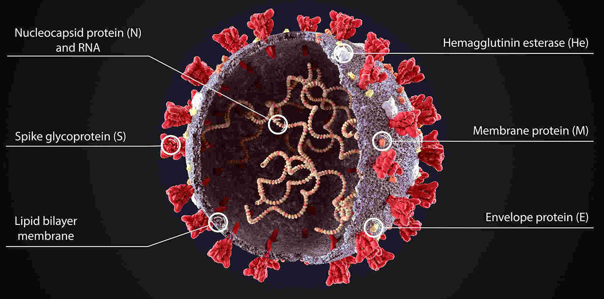
A class of single-stranded positive-sense RNA viruses known as coronaviruses have their name from the crown-like structure visible through electron microscopy which comes from their surface spike glycoproteins. RNA viruses include coronaviruses which possess one of the largest genomes that encode replicase along with structural proteins (S, E, M, N) and accessory proteins. Coronaviruses represent an ongoing global public health challenge because their rapid mutation ability allows them to transmit between different hosts.
| Genus | Subgenus/Lineage | Example Viruses | Host Range | Pathogenicity |
| Alpha-coronavirus | Traditional α-CoV | HCoV-229E, HCoV-NL63, PEDV, TGEV | Mammals | Humans: Common cold; Animals: Swine enteric diseases |
| Beta-coronavirus | A group (Embecovirus) | HCoV-OC43, MHV | Mammals | Humans: Common cold; Mice: Hepatitis and neurological damage |
| B group (Sarbecovirus) | SARS-CoV, SARS-CoV-2 | Humans, bats | Humans: Severe pneumonia, multi-organ failure | |
| C group (Merbecovirus) | MERS-CoV, Bat-CoV HKU4/5 | Humans, camels, bats | Humans: Severe pneumonia and renal failure | |
| D group (Hibecovirus) | Bat-CoV HKU9 | Bats | Animals: Asymptomatic or mild infection | |
| Gamma-coronavirus | No clear subgroups | IBV, Whale-CoV SW1 | Birds, marine mammals | Birds: Respiratory diseases; Marine mammals: Enteric infections |
| Delta-coronavirus | No subgroups | PDCoV, Avian-CoV | Pigs, chickens, ducks | Pigs: Enteric diseases; Birds: Mild respiratory symptoms |
Note: Porcine Epidemic Diarrhea Virus is the full name of PEDV while Transmissible Gastroenteritis Virus defines TGEV. IBV stands for Infectious Bronchitis Virus.
| Category | Cat. No. | Product Name | |
| Antibody | CABT-CS025 | Anti-Coronavirus OC-43 Nucleoprotein Monoclonal antibody | Inquiry |
| CABT-B343 | Anti-HCoV OC43 monoclonal antibody | Inquiry | |
| CABT-RM315 | Anti-HCoV HKU1 spike glycoprotein monoclonal antibody | Inquiry | |
| CABT-RM317 | Anti-HCoV spike glycoprotein polyclonal antibody | Inquiry | |
| ELISA Kits | DEIASL420 | Human anti-HCoV 229E IgM ELISA Kit | Inquiry |
| DEIASL423 | Human anti-HCoV NL63 IgM ELISA Kit | Inquiry | |
| DEIASL428 | Human anti-HCoV HKU1 IgG ELISA Kit | Inquiry | |
| DEIASL426 | Human anti-HCoV OC43 IgM ELISA Kit | Inquiry |
| SARS-CoV | MERS-CoV | SARS-CoV-2 | |
| Receptor and Invasion Mechanism | ACE2 receptor: S protein binds to ACE2 via RBD, TMPRSS2 protease cleaves to activate membrane fusion | DPP4 receptor: S protein binds to DPP4, primarily infects respiratory and renal epithelial cells | ACE2 receptor: S protein RBD binds to ACE2, Furin protease cleavage site (RRAR) enhances activation efficiency; TMPRSS2 or Furin cleavage promotes membrane fusion |
| Variation and Immune Evasion | Low variation rate, no significant immune evasion variants | Moderate variation rate, S protein L506F mutation enhances DPP4 binding stability | High variation rate: Delta variant increases transmissibility by 60%; Omicron variant shows significant immune evasion but reduced pathogenicity |
| Key Molecular Mechanisms | E protein activates NLRP3 inflammasome: induces IL-1β secretion, exacerbates lung inflammation | ORF4a inhibits interferon: blocks antiviral signaling by binding to MDA5 | ORF9b-Tom70 interaction: inhibits mitochondrial antiviral signaling protein (MAVS), weakens innate immune response |
| Disease Characteristics | Severe pneumonia with acute lung injury, cytokine storm leading to multi-organ failure | Severe pneumonia combined with renal failure, gastrointestinal symptoms | Primarily mild cases, severe manifestations include hypoxemia and thrombotic complications; "long COVID" sequelae (fatigue, cognitive impairment) are common |
| Pathogenic Stage | Viral Protein | Host Protein/Factor | Interaction Mechanism | Intervention Strategy |
| Viral Entry | Spike (RBD) | ACE2/DPP4 | Spike RBD binds to host receptor via polar residues, initiating membrane fusion | 1. Recombinant soluble ACE2 neutralizes the virus 2. RBD monoclonal antibodies block receptor binding |
| Membrane Fusion | Spike (S2 subunit) | TMPRSS2/Furin | TMPRSS2 or Furin cleaves the S1/S2 site of Spike, activating fusion peptide to drive viral-host membrane fusion | 1. TMPRSS2 inhibitors 2. 6-HB peptide (EK1) blocks fusion |
| Viral Replication | N protein, Non-structural proteins, Accessory proteins | G3BP1 + PI4K-IIIβ | N protein binds to G3BP1 to inhibit stress granule formation; PI4K-IIIβ generates PI4P to construct viral replication factories | 1. PI4K-IIIβ inhibitors disrupt replication factories 2. G3BP1 agonists restore stress granules |
| Immune Evasion | ORF8, ORF6, M protein | STAT1 + MHC-I | ORF6 binds to KPNA2 to block STAT1 nuclear entry; M protein inhibits STAT1 phosphorylation, interfering with Type I interferon signaling | 1. JAK-STAT activators restore interferon signaling 2. MHC-I agonists enhance antigen presentation |
| Inflammatory Damage | E protein, ORF3a | NLRP3 + NF-κB | E protein activates NLRP3 inflammasome to release IL-1β; ORF3a activates NF-κB pathway to induce IL-6/TNF-α secretion | 1. NLRP3 inhibitors 2. NF-κB inhibitors reduce cytokine storm |
Approved Vaccines and Technological Pathways
Part 01
mRNA Vaccines
Encode the S protein's RBD, inducing neutralizing antibodies and T-cell immunity.
Part 02
Adenovirus Vector Vaccines
Carry the S protein gene, activating both humoral and cellular immunity.
Part 03
Recombinant Protein Vaccines
Based on S protein trimer nanoparticles, combined with adjuvants to enhance immune responses.
Part 04
Inactivated Vaccines
Provide limited cross-protection against variants.
Peptide inhibitors designed to block membrane fusion in SARS-CoV-2, SARS-CoV, and MERS-CoV
S: Non-ACE2-BindingTargeting conserved epitopes outside the receptor-binding interface to reduce mutational escape risk
Broad-SpectrumCross-reactive immunity induced by M protein-specific T-cell epitopes
N ProteinHighly conserved nucleocapsid protein; combining N protein with RBD-based vaccines enhances the breadth of T-cell responses

New Products
Our antibodies serve mainly as research tools. Coronavirus antigen proteins detection and quantification through antibodies support scientific research on immune responses to the virus and enable the development of diagnostic kits and vaccines.
The recommended storage temperatures for antibodies are usually -20°C or 4°C and avoiding multiple freeze-thaw cycles ensures their activity and specificity remain intact. Despite antibodies not being typically biohazardous substances standard biosafety procedures should be implemented during handling to avoid contamination and accidental exposure.
To determine antibody specificityscientists must conduct cross-reactivity tests which compare antibody bindingto related viruses like SARS-CoV-1, MERS-CoV and cold coronaviruses toeliminate non-specific interactions. Western blot experiments require confirmationthat the observed band corresponds to the anticipated molecular weight of theintended protein. Under suitable conditions both knockdown and knockout of thetarget protein are possible to generate cell lines that serve as negativecontrols to validate antibody specificity.
You should confirm the following parameters:
The experimental applicability of antibodies depends on their epitope recognition characteristics and host species. Antibodies targeting linear epitopes (like those for the C-terminal peptide of N protein) are more suitable for Western Blot, while conformational epitope antibodies (such as those recognizing S protein trimers) are better for flow cytometry and live cell imaging. Cross-species reactivity needs to be confirmed through validation, for example, rabbit-derived antibodies used on mouse tissues require species absorption treatment.

Creative Diagnostics is a "One stop" antibody solutions provider serving the pharmaceutical, biotech, diagnostic and university research organizations around the world.
Tell us how we can support your project
Loading ......
