
 AAV Titration ELISA
Inquiry Basket
 Loading ......
The recombinant adeno-associated virus (AAV) vector belongs to the genus DNA dependoviruses of the family Parvoviridae. It is a small, non-enveloped DNA virus with a genome of approximately 4.7kb. In the past few years, AAV vector gene therapy has been used to permanently correct genetic diseases by copying the function of the gene into the nucleus of somatic cells, and has achieved certain success.
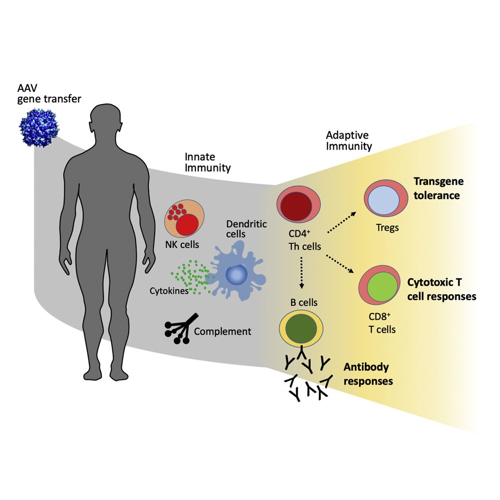 Figure 1. AAV vector immunogenicity in humans. (Verdera HC, et al.; 2020)
AAV vectors have unique advantages as gene transduction tools. For example, they are non-integrating, can transduce a variety of terminally differentiated tissues, drive long-term transgene expression, have low transduction efficiency in APC cells, and have low immunogenicity. Despite this, there is still a certain degree of immunogenicity when using AAV vectors for gene transduction, which may interfere with its effectiveness and cause safety concerns.
The immunogenicity of AAV vectors mainly comes from three aspects:
Seroepidemiological studies on human wild-type AAV neutralizing antibodies have shown that anti-AAV2 neutralizing antibodies have the highest detection rate, ranging from 30% to 60% of the population.
Due to the extensive cross-reactivity between AAV serotypes, neutralizing antibodies recognizing almost all serotypes can be found in subjects. The main reason is the amino acid sequence and structural homology between capsids of different AAV serotypes.
In addition to their neutralizing effects, antibodies have also been shown to increase accumulation of vector in lymph nodes. The presence of non-neutralizing antibodies increases AAV transduction in the liver. In addition, AAV antibodies may activate complement and cause toxic reactions.
In addition to humoral immunity, cellular immunity against AAV1 and AAV2 is also detected through technologies such as ELISPOT and flow cytometry. These AAV-specific T cells have high cross-reactivity for different serotypes and are more frequently found in the peripheral immune organs of the spleen than in peripheral blood. Most AAV-specific T cells have a memory phenotype and are found in adults and children. The detection level is close. It is possible that after AAV infection in infancy, memory phenotype-specific T cells are stored in peripheral lymphoid organs and persist throughout life.
AAV-specific memory T cells have been shown to produce IFN-γ, IL-2, and TNF-a and exhibit a cytotoxic phenotype characterized by granzyme B and CD107a degranulation markers.
AAV vectors lack sequences encoding viruses, and their main antigenicity comes from residual contaminants, viral capsids, and transduced gene products during the purification and production processes. The nucleic acid component of the viral vector may have an adjuvant-like effect, helping to activate the host immune system. Through pathogen-associated molecular patterns (PAMPs), antigens expressed in viral vectors can be recognized by PRRs (pattern recognition receptors) of immune cells, initiating natural immunity. These PRRs can recognize viral nucleic acids, as well as membrane glycoproteins and even chemical messengers. The involvement of PRR mainly leads to the activation of NF-kB and IRF transcription factors, which play a central role in inducing the expression of pro-inflammatory cytokines or type I IFN, respectively.
Adaptive immunity occurs after innate immunity through antigen-specific recognition, elimination of pathogens, and then establishment of immune memory. During the establishment of an adaptive response, T and B lymphocytes are activated upon recognition of antigens presented by APCs. Upon activation, lymphocytes expand and differentiate into effector cells and mediate elimination of antigen by inducing humoral or cytotoxic responses. After clearance of the antigen, the adaptive immune response is followed by a contraction phase that generates memory T and B lymphocytes that reactivate upon reexposure to the antigen. It has been demonstrated that genetically transfected cells and professional APCs present capsid protein epitopes to cytotoxic CD8+ T cells via MHC class I molecules. Cytotoxic T cells clear AAV-transduced cells, causing target organ inflammation and reducing the duration and effectiveness of gene transfer. Synchronous with MHC class II presentation, recognition of capsid-derived epitopes bound to MHC class II on the APC surface activates CD4+ T helper cells and promotes humoral and cell-mediated immune responses. Clinical trials have shown that AAV vector immunogenicity is dose-dependent to some extent, with low AAV vector doses more likely to cause mild inflammation without resulting in complete loss of transgene expression.
Similar to the capsid protein, the epitope of the gene transduction product is presented by APC, activating humoral and cellular immunity, and has a negative regulatory effect on the stability of AAV gene transduction. The key determinant of the level of immune response to the gene transduction product is the target organ, which is determined by the combination of the AAV capsid, vector delivery pathway, and tissue specificity of the promoter-driven gene expression. Systemic and intramuscular injections of vectors (using ubiquitous or muscle-specific promoters) are more immunogenic than injections into immune-privileged organs (using liver-specific promoters).
Anti-AAV Antibody ELISA Kit
AAV Antibodies and Titration ELISA
References
| Target | Cat. No. | Product Name | Size | Species Reactivity | Application | Detection Sample | |
| AAV1 | DEIASL342 | AAV9 Titration ELISA Kit | 96T | Qualitative | serum, plasma | Inquiry | |
| AAV2 | DEIASL347 | AAV6 Titration ELISA Kit | 96T | Human | Qualitative | Serum | Inquiry |
| DEIASL347M | Mouse Anti-AAV8 ELISA Kit(Quantitative) | 96T | Mouse | Qualitative | Serum | Inquiry | |
| AAV5 | DEIASL343 | AAV8 Titration ELISA Kit | 96T | Qualitative | serum, plasma | Inquiry | |
| AAV6 | DEIAAV6 | Anti-AAV2 antibody ELISA Kit | 96T | N/A | Quantitative | cell culture supernatants, purified virus preparations | Inquiry |
| DEIASL344 | Anti-AAV9 antibody ELISA Kit | 96T | Human | Qualitative | Serum | Inquiry | |
| DEIASL344M | Anti-AAV1 Antibody ELISA Kit | 96T | Mouse | Qualitative | Serum | Inquiry | |
| AAV8 | DEIASL345MQ | Anti-AAV5 Antibody ELISA Kit | 96T | Mouse | Quantitative | Serum | Inquiry |
| DEIAAV8 | Anti-AAV6 ELISA Kit | 96T | N/A | Quantitative | cell culture supernatants, purified virus preparations | Inquiry | |
| DEIASL345 | Anti-AAV8 antibody ELISA Kit | 96T | Human | Qualitative | Serum | Inquiry | |
| DEIASL345M | Mouse Anti-AAV9 antibody ELISA Kit | 96T | Mouse | Qualitative | Serum | Inquiry | |
| AAV9 | DEIAAV9 | Mouse Anti-AAV2 antibody ELISA Kit | 96T | N/A | Quantitative | cell culture supernatants, purified virus preparations | Inquiry |
| DEIASL348 | Mouse Anti-AAV6 antibody ELISA Kit | 96T | Human | Qualitative | Serum | Inquiry | |
| DEIASL348M | Mouse Anti-AAV8 antibody ELISA Kit | 96T | Mouse | Qualitative | Serum | Inquiry |
Loading ......
