






Inquiry Basket
 Loading ......
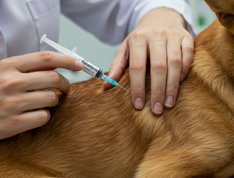
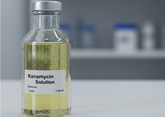
Kanamycin is a common broad-spectrum antibiotic. Kanamycin is used in laboratory research (such as molecular cloning screening and bacterial culture contamination control), clinical treatment, and veterinary fields. Do you want to know how Kanamycin is produced? How is it used in the above fields? And the problems and solutions brought about by its use? Creative Diagnostics has compiled a comprehensive Kanamycin information guide to ensure that researchers understand and use it correctly. Come and take a look.

Kanamycin functions as an aminoglycoside antibiotic with Kanamycin A serving as its principal component. Japanese scientist Hamao Umezawa isolated Kanamycin for the first time from Streptomyces kanamyceticus in 1957. Its chemical structure consists of three components: Kanamycin A represents the primary component with the highest proportion followed by B and C which stem from fermentation production. Kanamycin A's molecular structure includes 6-amino-6-deoxy-D-glucose and 3-amino-3-deoxy-D-glucose amino sugars which attach to 2-deoxystreptamine at its C4 and C6 positions through glycosidic bonds. The molecular formula for Kanamycin A is C18H36N4O11 and Kanamycin sulfate has a molecular weight of 582.58. The difference between Kanamycin B and C lies in the different linked sugar groups: The sugar groups in Kanamycin B include 2,6-diamino sugars whereas Kanamycin C contains 2-amino sugars.
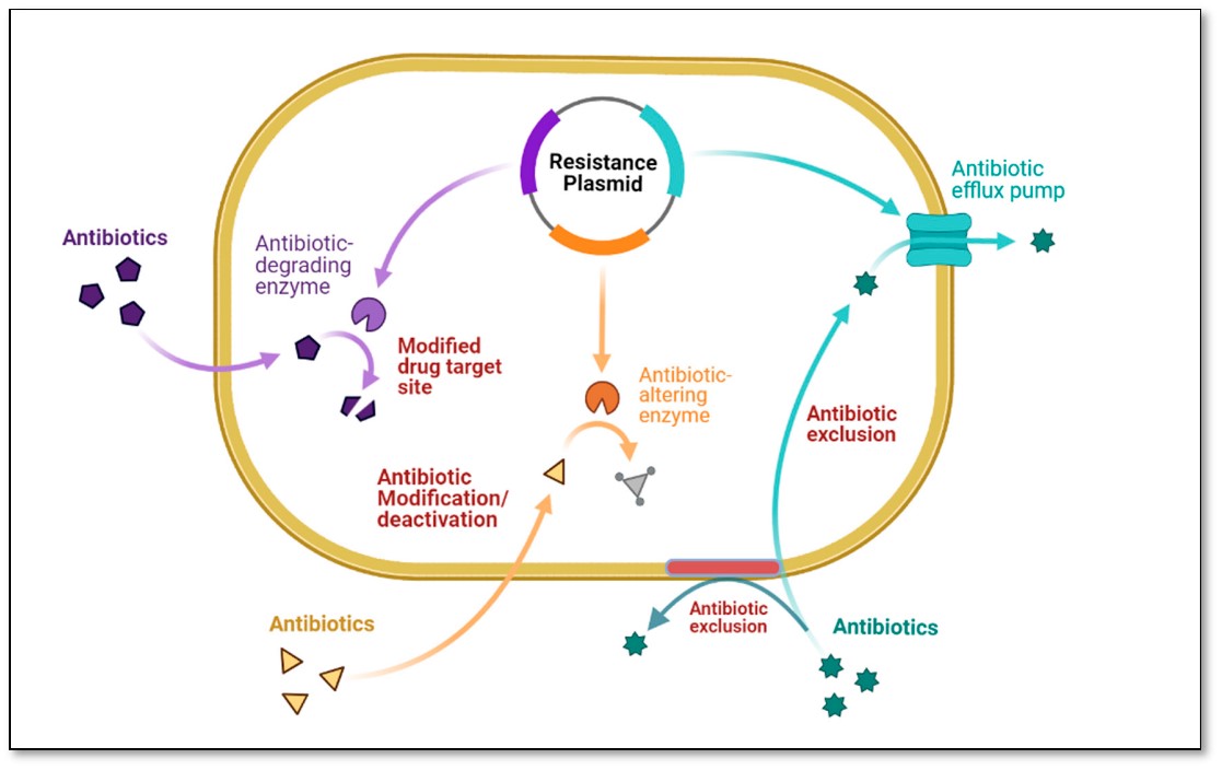
Kanamycin functions as an aminoglycoside antibiotic through its ability to target bacterial protein synthesis systems which leads to bactericidal effects by disrupting the translation process. Here we present an exhaustive analysis examining molecular biology aspects and structural binding interactions along with drug resistance mechanisms.
The bacterial ribosome comprises two distinct subunits known as the 50S and 30S subunits. The 30S subunit decodes mRNA sequences and binds initially with tRNA during protein synthesis. Kanamycin attaches itself to the 16S rRNA within the 30S subunit at a conserved area close to the A site where amino acid acyl-tRNA binds, particularly at the A1408 and G1491 bases of the 16S rRNA. The deoxystreptamine ring and amino sugar group attach to the phosphate backbone of 16S rRNA using hydrogen bonds and electrostatic interactions which change the 30S subunit structure and disrupt its accurate pairing with mRNA and tRNA.

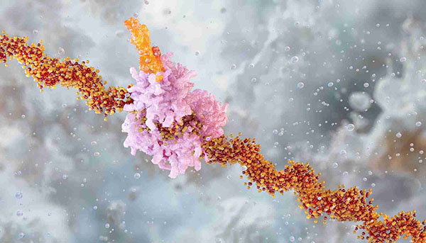
The antibiotic kanamycin disrupts translation by affecting all three stages which include initiation, elongation and termination. During initiation stage translation, it blocks the assembly of the initiation complex (30S - mRNA - fMet - tRNA) and disrupts the detection of the Shine - Dalgarno sequence with the 30S subunit which stops translation from starting. The elongation stage encounters two major disruptions from the agent which prevents proper codon recognition by the ribosome leading to insertion errors like misreading leucine as isoleucine and simultaneously blocks the A site function preventing aminoacyl-tRNA entry and hindering tRNA release thus creating an inactive ribosome. The translation termination stage disruption prevents termination codon recognition (UAA, UAG, UGA) which leads to ribosome stalling on mRNA and results in non-functional polyribosome formation.
The bactericidal activity of Kanamycin not only relies on translation inhibition, but is also enhanced through two other pathways. The compound starts by breaking down the cell membrane integrity. Membrane potential collapse occurs because improper protein incorporation leads to cell membrane pores that permit K+ and Mg2+ ion leakage. By binding to bacterial outer membrane lipopolysaccharide (LPS) the substance increases membrane permeability which allows drug entry through a self-enhanced uptake process. The compound activates bacterial production of reactive oxygen species that cause DNA damage and energy resource depletion.

| Comparison Dimension | Kanamycin | Gentamicin | Amikacin | Tobramycin | Streptomycin | Etimicin |
| Antimicrobial Spectrum | Gram-negative bacteria (E. coli, Klebsiella), some Staphylococci, M. tuberculosis (second-line) | Gram-negative bacteria (P. aeruginosa, Enterobacteriaceae), some Staphylococci | Broad Gram-negative coverage (including gentamicin-resistant strains), P. aeruginosa | Gram-negative bacteria (highest activity against P. aeruginosa) | Gram-negative bacteria, M. tuberculosis (first-line), Y. pestis | Gram-negative bacteria (respiratory/urinary infections) |
| Resistance Mechanisms | Inactivated by APH(3')-I enzymes | Susceptible to 6 types of modifying enzymes (high cross-resistance) | Resistant to most modifying enzymes | Similar to gentamicin but more stable against some enzymes | Relies on rpsL mutations (in M. tuberculosis) | Similar to gentamicin (lower nephrotoxicity) |
| Pharmacokinetics | IM peak: 1 hr; half-life: 2.5 hr; renal excretion | Half-life: 2 hr; renal excretion (requires TDM) | Half-life: 2.5 hr; superior tissue penetration | Half-life: 2-3 hr; high lung concentration | Half-life: 5 hr; penetrates tuberculous lesions | Half-life: 3 hr; low nephrotoxicity |
| Clinical Use | MDR-TB, intra-abdominal infections | Sepsis, pneumonia, complex UTIs | Multidrug-resistant Gram-negative infections, HAP | P. aeruginosa infections (cystic fibrosis) | Tuberculosis (first-line), plague | Respiratory/UTIs (China-specific) |
| Ototoxicity/Nephrotoxicity | High ototoxicity (20-60% cumulative risk), moderate nephrotoxicity | High vestibular toxicity, nephrotoxicity | Lower toxicity vs. gentamicin | Lower toxicity vs. gentamicin | Auditory toxicity, moderate nephrotoxicity | Low nephrotoxicity, moderate ototoxicity |
| Neuromuscular Blockage Risk | Moderate risk (competes with Ca²⁺) | Low risk (caution with anesthetics) | Low risk | Low risk | High risk (avoid with muscle relaxants) | Low risk |
| Cross-Resistance | Partial with gentamicin/tobramycin | High with kanamycin/tobramycin | Effective against gentamicin-resistant strains | High with gentamicin | No cross-resistance | Partial with gentamicin |
| Unique Advantages | Core drug for MDR-TB | Cost-effective, broad-spectrum | Enzyme-resistant, first-line for MDR strains | Best anti-Pseudomonas activity | Irreplaceable for TB/plague | China-developed, optimized safety |

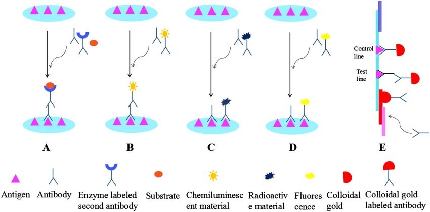
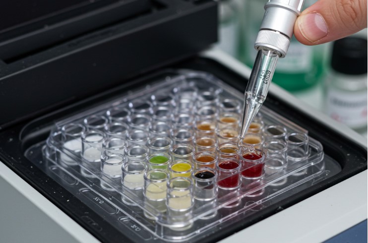
The Kanamycin ELISA test operates through competitive binding principles. The enzyme-labeled antibody binds with both pre-coated antigen and kanamycin from the sample through competition and enables quantitative detection of the leftover amount through a colorimetric reaction. The detection range extends from 0.5 ng/mL up to 40.5 ng/mL while sensitivity reaches a level of 0.1 ppb. Residue detection technology finds widespread application in both food safety testing (including milk and meat) and biopharmaceutical analysis (including vaccines and plasmid DNA). Complex samples need to be processed by TCA precipitation or dilution to reduce matrix interference. Rapid detection technologies such as colloidal gold test strips can complete on-site screening (such as raw milk) within 5-10 minutes, with a detection limit of 20 μg/kg. The focus of kit development is on antibody specificity optimization and standardized production to meet pharmacopoeia and industrial needs.
Low cost per test: Compared with HPLC or LC-MS/MS, it does not require expensive instruments and maintenance costs, and is suitable for large-s cale screening
| Target | Cat. No. | Product Name | Host | Isotype | Application | |
| Kanamycin | HMABPY044 | RHA™ anti-Kanamycin Monoclonal antibody, clone KAN | Mouse | IgG | ELISA, LFIA | Inquiry |
| Kanamycin | DPABY-883 | Anti-Kanamycin polyclonal antibody | Sheep | ELISA, Pr* | Inquiry | |
| Kanamycin | DPAB-DC4461 | Anti-Kanamycin polyclonal antibody | Sheep | EIA | Inquiry |
| Target | Cat. No. | Product Name | Expression System | Tag/Conjugate | Application | |
| Kanamycin | DAG1208 | Kanamycin [HRP] | N/A | HRP | N/A | Inquiry |
| Kanamycin | DISNJ15 | Kanamycin Sulfate Standard | N/A | N/A | ELISA | Inquiry |
| Kanamycin | DAGA-040B | Kanamycin [BSA] | N/A | BSA | LFIA | Inquiry |
| Kanamycin | DAGA-044K | Kanamycin [KLH] | N/A | KLH | Immunogen | Inquiry |
| Kanamycin | DAG227S | Kanamycin [HSA] | N/A | HSA | ELISA | Inquiry |
| Kanamycin | DAG515S | Kanamycin [HSA-Biotin] | N/A | HSA-Biotin | ELISA | Inquiry |
| Target | Cat. No. | Product Name | Size | Species Reactivity | Application | Detection Sample | |
| Kanamycin | DEIAH-004H | Kanamycin ELISA Kit | 96T | Quantitative | milk, milk powder, chicken, pork | Inquiry | |
| Kanamycin | DEIA048 | Kanamycin ELISA Kit | 96T | N/A | Quantitative | biological samples | Inquiry |
| Kanamycin | DEIA-WZ048V | High Sensitivity Kanamycin ELISA Test Kit | 96T | Quantitative | biological samples | Inquiry | |
| Kanamycin | DEIA048V | Kanamycin ELISA Kit | 96T | Quantitative | Vaccine | Inquiry | |
| Kanamycin | DEIA-004H | Kanamycin ELISA Kit | 96T | N/A | Quantitative, qualitative | tissue, milk | Inquiry |
| kanamycin | DTS761 | Kanamycin Residue Rapid Test(milk) | 96T | N/A | Qualitative | Milk | Inquiry |
Kanamycin is an aminoglycoside antibiotic and is widely used in scientific research, especially in molecular biology and biochemical research. Its main scientific research applications include the following aspects:
Gene Screening and Cloning
Kanamycin is often used to screen clones carrying kanamycin resistance genes. Its resistance gene is usually used as a marker gene to screen successfully transformed cell lines through antibiotic selection pressure.
Microbial Culture and Antimicrobial Activity Testing
The culture medium uses kanamycin solution as an antibiotic to prevent non-target microorganism growth which ensures target strain purity during experimentation. LB agar plates receive kanamycin during culture processes to select for kanamycin-resistant bacteria.
ELISA Detection
Researchers use kanamycin to create different ELISA kits capable of detecting kanamycin residues present in samples. The available kits are composed of indirect competitive ELISA and competitive ELISA methods which show high sensitivity and accuracy for detecting kanamycin residues in food products, animal feed as well as biopharmaceuticals.
Chemiluminescent Immunoassay
Scientists use kanamycin monoclonal antibodies to create chemiluminescent immunoassay kits which detect kanamycin residues found in milk and milk powder. This detection method is rapid and sensitive.
Biochemical Analysis and Experimental Interference Studies
Kanamycin solution is also used as an interfering substance in biochemical experiments to evaluate the impact of experimental conditions on test results. For example, researchers test the degree of interference with experimental results by adding different concentrations of kanamycin.
Radiolabeling and Clinical Diagnosis
Kanamycin is also used in radiolabeling studies, such as the preparation of 99mTc-kanamycin for early infection detection.
Kanamycin ELISA detection sensitivity optimization requires enhanced monoclonal antibody affinity together with precise antigen epitope design. The antibody specificity gets much better when you find the right ratio to couple kanamycin with carrier proteins like BSA. The reduction of cross-reactivity requires the selection of highly specific antibodies through screening. Research indicates that kanamycin assay kits demonstrate cross-reactivity rates lower than 0.1% when tested against gentamicin and neomycin. The detection limit can be reduced to below ppb level when nanomaterials like colloidal gold or fluorescent labeling technology are introduced.
The interference of complex samples can be solved by the following methods:
Immunogen design: Use EDC method to couple kanamycin to carrier protein (such as BSA) to ensure epitope exposure.
Difficulty: Rapid detection needs to balance speed (5-10 minutes) and accuracy to avoid false positives/false negatives.
Solution: Use double antibody sandwich method to optimize color development logic and improve color development stability.
Loading ......
