




Inquiry Basket
 Loading ......
What aspect of the specific application of the antibody would you like to inquire about?
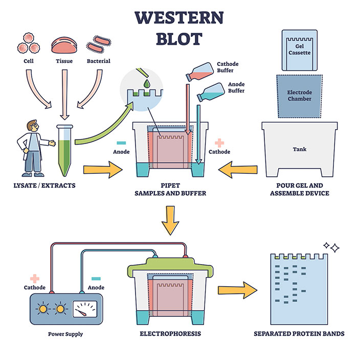
This issue is often related to transfer efficiency, antibody concentration, or sample quality. Start by verifying whether the protein transfer was successful. Even if the transfer process is correct, then the issue may be low antibody level or damaged antibodies. For that reason, increase the antibody level or make new antibodies. Also, you can try loading additional sample or changing the lysis buffer composition if target protein isn’t expressed to increase protein removal rate.
Excessive background may result from insufficient blocking, overly high antibody concentrations, or inadequate washing. To address this, consider extending the blocking duration and using fresh blocking solutions, such as 5% non-fat milk or BSA, while ensuring that the blocking agent does not react with the antibodies. Then, if that still isn’t working, reduce the primary and secondary antibodies and up the washing speed and time. Further, longer exposure times can be used to make the background noise appear stronger, and should therefore be minimized if you can.
Nonspecific bands may arise from insufficient antibody specificity or sample degradation. To minimize nonspecific binding, it is recommended to use monoclonal antibodies or affinity-purified polyclonal antibodies. During sample preparation, ensure the addition of protease inhibitors to prevent protein degradation. If these factors are not the issue, consider reducing the sample loading amount, as excessive protein can lead to nonspecific band formation.
Bad storage (freeze-thaw cycles, for example, are the most common causes of antibody degradation). To avoid this, aliquot the antibody and store it at -20°C or -80°C, minimizing freeze-thaw cycles. For working solutions, prepare them fresh before use and avoid prolonged storage. Also shield antibodies from intense light, as some fluorescent secondary antibodies are light sensitive and will not be active when in contact with light in some experiments.
Spotted background is often caused by particulate impurities in the blocking solution or aggregation of the secondary antibody. To address this, filter both the blocking solution and the secondary antibody to remove any particles. Additionally, ensure that the incubation solutions are well-mixed during the process and avoid using reagents that have precipitated. You can optimize each step, and adjust experimental conditions as per the specific problems, and the success and data quality of Western Blot experiments can greatly improve. If it continues to malfunction, follow the manufacturer’s advice or ask technical support for further information.
If you want to get more antibody specificity in WB, here are a few tips:
Use tested-and-proven antibodies. Choose your antibody depending on the need. Monoclonal and affinity-purified polyclonal antibodies are generally more specific. The recombinant monoclonal antibodies, optimized using genetic engineering, are more specific and reproducible.
Improving specificity often involves fine-tuning experimental parameters. Using the proper blocking agent (BSA or nonfat milk) to reduce nonspecific binding and selecting the correct buffer for primary antibody dilution as specified by the manufacturer. Set the primary antibody incubation parameters (temp and time) to minimize nonspecific binding and maximize specific binding.
Overloading protein samples can lead to nonspecific bands. Reduce the amount of sample loaded to improve band clarity. Ensure proper transfer conditions—proteins should be fully transferred but not excessively so, to avoid loss of target proteins or excessive background. Additionally, select the appropriate gel concentration based on the size of your target protein to achieve optimal separation.
Ensure that antibody levels are properly stored. Conserve antibodies at recommended storage temperatures (-20°C or -80°C) and do not freeze-thaw them in the same batch again, by aliquoting. Prep new working solutions of primary antibodies for use and do not store dilute solutions for long periods of time as this will degrade or cause contamination.
If you take these steps and work hard on optimizing every step of your protocol, then antibodies will be significantly more specific in WB experiments, and the data will be more consistent and precise.
You will have to choose the right antibody for IHC to be successful. Be sure the antibody is confirmed for IHC experiments and especially for detection of the target protein in its native form.
You can make your selection through the following steps:
Weak or absent signals may result from the following reasons:
Nonspecific staining could be caused by one of the following factors:
Inconsistent staining results may be due to suboptimal experimental conditions. You can use positive and negative control tissues to validate the workflow. Also, be sure to do everything exactly the same in different experiments, and make sure the reagents are expiring and not contaminated.

More Information about Antibodies for Immunohistochemistry (IHC)
Use Appropriate Controls:
Assess Antibody Specificity:
Cross-Validation with Other Techniques:
Optimizing Primary Antibody Incubation Time:
Commonly recommended incubation times include short-term incubation at room temperature (RT) and long-term incubation at low temperatures (4°C). For low-abundance targets, overnight incubation can enhance signal intensity.
The incubation time is influenced by both the antibody concentration and the target protein's abundance. During preliminary experiments, test various incubation durations and evaluate changes in the signal-to-noise ratio. If background staining is high, consider shortening the incubation time or reducing the primary antibody concentration.
Optimizing secondary antibody incubation time:
Secondary antibodies are typically incubated at room temperature for 30 minutes to 1 hour. Some commercial kits may suggest even shorter incubation times.
Be careful not to prolong secondary antibody incubation as it might lead to extra nonspecific binding. Moreover, with fluorescent secondary antibodies, keep incubation in a dark room to avoid photobleaching the fluorescent signal.
Balancing temperature and incubation time:
Short-term incubation at room temperature is suitable for rapid experiments but may result in higher background signals. Low-temperature, long-term incubation (e.g., overnight at 4°C) improves specificity, particularly for high-affinity antibodies or complex samples.
If a shorter experimental timeline is required, increasing the antibody concentration at room temperature can compensate for reduced incubation time. However, this approach should be carefully optimized to avoid excessive nonspecific binding.
Estimate Antibody Binding Efficiency Based on Fluorescent Signal Strength:
Perform Quantitative Detection with Flow Cytometry:
Set Up Appropriate Controls:
More Information about Antibodies for Immunocytochemistry (ICC)
| Possible issues | Possible causes | Solutions |
| Weak or No Signal | Possible causes include inappropriate fixation, incorrect antibody dilution, or insufficient incubation time | Solutions involve optimizing fixation protocols, adjusting antibody concentrations, and ensuring proper incubation conditions |
| High Background | High background staining can result from insufficient blocking, non-specific binding of antibodies, or autofluorescence from samples | To reduce background, increase blocking incubation time, use appropriate blocking agents, and ensure thorough washing between steps |
| Non-Specific Staining | The antibodies might bind outside the target but not within its bound area (i.e., cross-reactivity) | Runnable controls (e.g., isotype controls) can flag general binding problems |
Signal diminution is a common issue in IF experiments that can affect the reliability of results. Here are some effective strategies and recommendations to reduce signal loss and enhance the success of your experiments.
| Directly Labeled Primary Antibodies | Indirectly Labeled Secondary Antibodies | |
| Advantages | Single-Step Method: Direct immunofluorescence is performed using a first antibody that is directly attached to a fluorophore. This ensures an easy staining process with the antibody reacting directly to the antigen target (without the secondary antibody) | Signal Amplification: It will be signal amplification because multiple secondary antibodies can bind to each primary antibody. It can therefore be highly sensitive and is therefore suitable for low abundance targets |
| Speed and Simplicity: This method is generally faster and simpler, as it requires fewer steps and reagents. It can reduce background noise and minimize issues related to cross-reactivity from secondary antibodies. | Flexibility and Multiplexing: Indirect methods offer greater flexibility in experimental design. A single secondary antibody can be used with various primary antibodies from different species, facilitating multiplexing in experiments | |
| Disadvantages | Lower Sensitivity: The directly labeled antibodies can be less sensitive than the indirect ones. There are only so many fluorophores that can be attached to one primary antibody, which is a limitation when looking for low-yielding proteins | Complexity and Time Consumption: The indirect approach is generally more complex and time-consuming due to the additional steps involved. It may also introduce background noise and increase the risk of cross-reactivity. |
| Minimized Multiplexing Potential: Direct labelling could prevent several antibodies of the same host species being able to be employed at once since each directly labeled antibody will send only one signal |

More Information about Antibodies for Immunofluorescence (IF)

Avoid IgG overlap signal problems with WB by using antibodies from different species during IP and WB. Furthermore, using the right secondary antibodies can reduce the noise.
(1) Select Appropriate Antibodies
(2) Optimize Incubation Conditions
(3) Adjust Washing Conditions
(4) Use Crosslinking Agents
In some cases, antigen-antibody complexes can be stabilized with crosslinking agents. This is particularly effective for proteins with weak interactions.
(5) Pre-purify Samples
Before conducting IP, pre-treating the lysate with similar components can help reduce impurities in the lysate, thereby enhancing both the recovery rate and binding efficiency of the target protein.
More Information about Antibodies for Immunoprecipitation (IP)
For better odds of success, choose antibodies that have already been tested for ChIP or something like that. It’s also helpful to make sure the epitope the antibody recognizes is present following crosslinking and fragmentation of chromatin.
Positive controls are antibodies that are already able to recognize target DNA. Positive controls include antibodies to histone H3 or RNA polymerase II (RNA Pol II), for instance. Such antibodies ensure the bound protein appears where it should in a genomic area and so verify the success of the experiment. If the positive control shows the anticipated signal, it indicates that the chromatin structure and experimental conditions are suitable.
Negative controls typically involve using normal IgG antibodies, which should not bind to any specific epitopes. By comparing the signals from the negative control samples with those from the target samples, one can assess the extent of non-specific binding. If the signal detected in the negative control samples is similar to that in the target samples, it may suggest the presence of non-specific binding or excessive background signals.
When conducting ChIP experiments, it is essential to include both positive and negative controls for each sample. This not only allows us to test whether the antibodies are specific or not but it also eliminates any risks of mishaps during the experiment. When results are pooled in samples, it is easier to discern how the target protein actually attaches in which areas.
During the qPCR phase, primers should be designed for genomic loci known to either bind or not bind the target protein, serving as positive and negative controls. These primers can help confirm whether ChIP results are reliable and provide further data support.
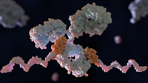
More Information about Antibodies for Chromatin Immunoprecipitation (ChIP)

FC works with monoclonal and polyclonal antibodies. mAbs are more specific, but pAbs can be more sensitive because they recognize more than one epitope. One should look for antibodies that are proven to work on FC.
You have to titrate antibodies when they’re new in the experimental setting, when cell types are changed, or when you switch fluorochromes. A titration series will usually have several different antibodies tested at different concentrations, to determine the best dilution and signal-to-noise ratio. This process should be performed on the same cell type intended for staining in the actual experiment.
Choose fluorochromes that are bright, have a spread in spectral distribution and overlap with the other fluorochromes in your panel. Lighter fluorochromes are good for more obscure targets, but these can improve sensitivity.
Also, always include positive and negative controls. Positive controls make sure your staining is going as planned, and negative controls filter out errant signals and unspecific binding.
Be sure to use positive and negative controls in your experiments. Positive controls check if your staining is accurate, and negative controls block unwanted signals and non-specific binding.
ELISA typically employs two antibodies – primary antibodies which recognize the antigen, and secondary antibodies that attach to the primary antibody. Secondary antibodies will be paired with enzymes or other label that is detectable.
For selecting ELISA antibodies, think of how highly sensitive the antibody is to the antigen. Be also sure that the secondary antibody corresponds to the species in which the primary antibody was generated. Cross-reactivity is reduced and assay efficiency improved by using the same antibody pairs.
To optimize an ELISA protocol, store all reagents in fresh containers. Split up primary and secondary antibodies to the correct concentrations. Additionally, follow a consistent washing protocol to remove unbound antibodies effectively.
Common troubleshooting steps include:

More Information about Antibodies for Immunoassays and ELISA
Loading ......
