




Inquiry Basket
 Loading ......
Antibodies are fundamental experimental agents in protein research. Most laboratory tests, WB, IP, IHC, IF, ChIP, and FC, all use antibodies. And how do you choose the right antibody for your experiment, and apply it effectively? Creative Diagnostics has written a comprehensive antibody selection and usage guide to make sure that researchers are using it the right way. Come and have a look.


When selecting an antibody, the first thing to consider is antibody specificity. The selection of specificity mainly needs to consider four aspects: protein specificity, species specificity, experimental method specificity, and marker specificity.
When searching for antibodies for the protein to be detected, several details should be distinguished:
a. If the recombinant protein is not expressed in full length, it is necessary to pay attention to whether the immunogenic region of the antibody is within the recombinant protein region.
b. It is best to know the way of shearing and modification of endogenous proteins. Proteins with special phenotypes need to be sequenced and combined with the antibody immunogenic sequence to check the cross-reaction.
c. Phosphorylated protein detection requires the determination of specific sites. Phosphorylation at different sites means that there may be different mechanisms, which should not be generalized.
The same protein has different species, which are larger or smaller.
a. At present, most commercial antibodies are recombinant proteins or designed polypeptide antigens based on human protein sequences as templates, and cross-react with other species according to the homology of the protein. It is necessary to refer to the reactive species information indicated in the instructions.
b. It is difficult to find antibodies for some rare species. You can select antibodies with homologous sequence immunity by comparing the protein sequence.
There are many experimental methods using antibodies at present. In different methods of use, because the processing methods of protein samples are different, the protein content is different, and the requirements for antibody recognition epitopes and titers are different.
Generally, experimental methods based on experimental operations will not use labeled primary antibodies, such as WB, IHC, etc., which are all labeled by secondary antibody reagents to achieve the purpose of presenting results. However, some experiments based on instrument analysis may use directly labeled primary antibodies, such as flow cytometry experiments. Then you need to understand the fluorescence range that the instrument you are going to use can detect, and select the corresponding markers for different parameter requirements. In immunofluorescence double labeling experiments, different fluorescent markers need to be selected.
| Category | Application | Description |
| Research | Western Blotting | Detect specific proteins in a sample using antibodies specific to the target protein. |
| ELISA (Enzyme-Linked Immunosorbent Assay) | Quantify proteins, peptides, antibodies, and hormones in samples. | |
| Immunohistochemistry (IHC) | Detect and visualize specific antigens in tissue sections. | |
| Immunofluorescence (IF) | Detect antigens in cells or tissues using fluorescent-labeled antibodies. | |
| Flow Cytometry | Analyze cell populations and quantify specific cell surface or intracellular molecules. | |
| Immunoprecipitation (IP) | Isolate and concentrate a particular antigen from a sample using a specific antibody. | |
| Diagnostic | Diagnostic Immunoassays | Detect biomarkers for diseases, such as infectious diseases, cancers, and autoimmune disorders. |
| Point-of-Care Testing | Rapid diagnostic tests for immediate results at the site of patient care. | |
| Immunohistochemistry (Diagnostic) | Diagnose diseases by detecting specific antigens in tissue samples. | |
| Therapeutic | Monoclonal Antibody Therapy | Treat diseases such as cancers, autoimmune diseases, and infectious diseases. |
| Antibody-Drug Conjugates (ADCs) | Targeted cancer therapy combining antibodies with cytotoxic drugs. | |
| Immune Checkpoint Inhibitors | Block proteins that prevent the immune system from attacking cancer cells. | |
| Prophylactic | Vaccines | Prevent infectious diseases by stimulating an immune response using antigens or antibodies. |
| Analytical | Biosensors | Detect specific biological molecules using antibody-based recognition elements. |
| Surface Plasmon Resonance (SPR) | Measure interactions between biomolecules in real-time without labeling. | |
| Biolayer Interferometry (BLI) | Analyze biomolecular interactions in real-time. | |
| Functional Studies | Blocking/Neutralizing Assays | Inhibit the activity of specific proteins or receptors to study their function. |
| Apoptosis Detection | Identify and quantify apoptotic cells using antibodies against specific apoptotic markers. | |
| Quality Control | Contaminant Detection | Detect and quantify contaminants such as host cell proteins and DNA in biopharmaceutical products. |
| Stability Testing | Assess the stability and shelf-life of biopharmaceutical products using specific antibodies. | |
| Personalized Medicine | Companion Diagnostics | Select appropriate therapies based on the presence of specific biomarkers detected by antibodies. |
We first need to know the type of antibodies required for these experiments in order to understand the difference in the antibody requirement in above techniques.
Once boiled and denaturated, WB proteins are linear. So the antibodies best are experiments on artificially made peptides with highly defined sequences, and the results are highly defined too.
For IP, antibodies derived from purified natural proteins or antibodies derived from purified recombinant proteins be used. Don’t use antibodies produced from peptides made artificially, since the sites of antibodies might be hidden deeply in the protein’s centre. The CHIP isn’t so different from IP. The catch is, if the epitope that the antibody recognizes matches the place where the protein latches on to DNA, the CHIP experiment will go awry.
Both immunofluorescence and immunohistochemistry need a fixation step, a determination of whether the cells’ morphology will remain consistent with the original. The denatured, coagulated protein produced by this chemical fixation is a bit unlike the normal protein but it is not the linear structure destroyed when WB heats it. So for IF/IHC, the best antibodies to use are antibodies isolated from purified recombinant proteins or antibodies isolated from synthetic peptides (peptides reside on the surface of proteins)
The flow cytometry is either the flow cytometry of live cells, best with antibodies against natural proteins or recombinant proteins, or the flow cytometry after fixation, best with the antibodies in IF/IHC. In flow cytometry, we have direct labeling and indirect labelling and the indirect labelling isn’t as good as the first. So we pick fluorescing flow cytometry antibodies, if the protein in question doesn’t already have a fluorescently labeled antibody, an indirect labeled antibody is selected and then a fluorescent secondary antibody is added.
You get more than one antibody for any target protein detection. To limit the antibody choice and choose the right antibody, here are some things to consider:

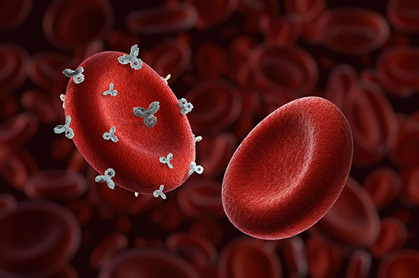

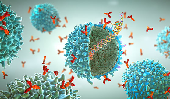
An antibody called a monoclonal antibody is an antibody generated by a clone. Monoclonal antibodies could interact only with one antigenic determinant. Antibodies raised against more than one antigenic determinant.
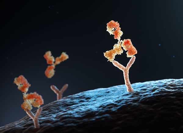
Secondary antibodies are - very broadly - antibodies that act specifically and bind to primary antibodies. There is more than one antibody to look for for any target protein. When choosing secondary antibodies, primary antibody and the needs of later detection algorithms should be taken into account. In general, the choice of the right secondary antibodies must be approached from these angles:
Select the species secondary antibody according to the species of the primary antibody.
The second antibody should belong to the same class or subclass as the first. That is usually monoclonal antibodies. Polyclonal antibodies consist mostly of IgG immunoglobulins, so second antibody is anti-IgG antibody. Class and subtype of monoclonal antibodies usually mentioned in the manual. If the former antibody is mouse IgM, then it should be anti-IgM. If the monoclonal first antibody is an IgG1 sub-type of mouse IgG (IgG2a, IgG2b, IgG3), virtually any anti-mouse IgG can be attached to it, or you can designate a subtype-specific secondary antibody. This antibody class can be especially used for double labeling.
For the most part, probes attached to the secondary antibody are enzymes (horseradish peroxidase HRP), fluorescent proteins, biotin and gold particles. Which secondary antibody goes with which probe is largely up to experiment. The WB and ELISA secondary antibodies that are used are enzyme-labeled secondary antibodies; fluorescent group-labeled secondary antibodies tend to be used in immunofluorescence experiments, and gold particle-labeled secondary antibodies are used mainly in immunoelectron microscopy.
| Category | Guidelines |
| Dilution and Incubation of Antibodies |
|
| Storage and Handling of Antibodies |
|
| Control Conditions in Experiments |
|
| Record and Report |
|
| Experimental Optimization |
|
Not necessarily. Is direct or indirect labeling applied to flow cytometry? If it is direct labeling then typically the antibody is not FITC or TRITC or PE etc. If you happen to need fluorescent labelled antibodies to direct label for immunohistochemistry, then it can be used. If your immunohistochemistry involves other labels, or indirect labeling, you’re in trouble, because the fluorescent labeling affects the bound to the secondary antibody and the primary antibody.
Your antibody may produce by artificially synthesizing peptides. The peptide you apply as an antigen is somehow stuck in the middle of the protein so its linear portion can be detected by WB.
Obviously, no! Immunofluorescence analysis uses immunofluorescent antibodies. They are FITC or PE, etc. labeled antibodies. The output has to be excited by UV light and read under a fluorescence microscope. WB antibodies are generally HRP or alkaline phosphatase, etc., antibodies that demand color substrates (DPTMB, etc.) to produce color directly (naked eye) or chemiluminescence (you need special tools).
Not necessarily. Antibodies in the CHIP class generally have rather high standards for purity and specificity. You can really say that antibodies that you can use for CHIP, you can use for WB. But if the CHIP antibody recognizes a conformational epitope (ie, two ends that are really very close together but much away after it becomes a linear structure) rather than a linear epitope, then this CHIP antibody is not capable of WB.
Dilution for WB antibodies: 1:1000, for IF/IHC: 1:100, and for ELISA, if possible: 1:10000. If the antibody is for ELISA then the idea is the antibody titer is not high. But that does not mean that WB cannot be performed with ELISA antibodies. The antibody manual says 1:1000 for ELISA, you can try 1:100 for WB.
Loading ......
