
Inquiry Basket
 Loading ......
Antibodies are key proteins that play a central role in the immune system, specifically as affinity proteins produced by B cells. Antibodies exhibit extremely high specificity and affinity, enabling them to lock onto unique molecular markers on pathogens' surfaces (antigen epitopes) and bind to specific antigens. This binding triggers a series of immune responses such as phagocytosis, lysis, and memory immunity. As the core molecules of humoral immunity, antibodies form the foundation of adaptive immunity and are nature's "precision weapons" in biology. They function like "guided missiles," accurately identifying and neutralizing invading foreign pathogens. Antibodies are not only defense tools for the body but also serve as central molecules for coordinating and regulating complex immune reactions, with their role permeating every stage of the immune response.
Antibodies are Y-shaped macromolecules composed of two heavy chains (H chains) and two light chains (L chains), each part serving a specific task: from antigen recognition to immune response activation, precisely coordinating and executing the antibody's defensive functions
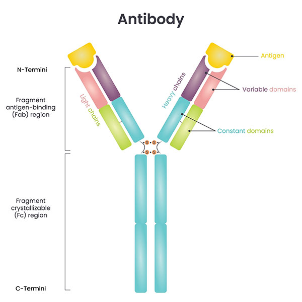
The immunoglobulin fold (Ig-fold) is formed by two tightly packed, anti-parallel β-folds, resulting in a compact structure. Each chain (heavy and light) contains around 110 amino acid residues. β-strand stacking forms each domain, creating a "β-barrel" structure. Disulfide bonds connect these domains and stabilize the overall antibody structure.
The Fab fragment, located at the top of the Y-shaped antibody molecule, is responsible for antigen specific recognition. Each Fab consists of the following components:
The hinge region is located between the Fab and Fc fragments and provides flexibility and movement for the antibody. This flexible "rope" allows the Fab fragments to adjust their angles according to the shape of the antigen without destabilizing the antibody. By altering the relative angle between the Fab and Fc, the two Fab arms can independently bind to different antigens. In fact, the elbow angle of the Fab can range from 116° to 226°, providing a wide range to accommodate different antigen shapes. This flexibility ensures that antibodies can function more efficiently in complex immune environments.
The Fc fragment is the antibody’s "effector function region" that is not engaged in antigen recognition, but stimulates immune activity through interaction with the rest of the immune system (immune cell receptors, the complement system).
Glycosylated Fc: The Fc region contains glycosylation alterations that serve as "switches" to regulate antibody function. Glycosylation affects both antibody stability and receptor binding capacity.
Attachment to Immune Cell Receptors: Fc attaches to Fc receptors, causing ADCC, ADCP, and other immune activations. It improves the function of immune cells including macrophages and NK cells.
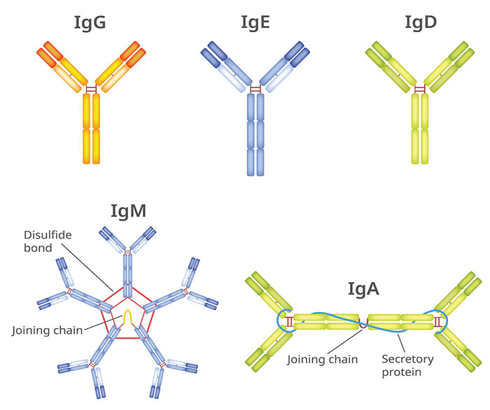
IgG: As the most adaptable antibody in the family, IgG helps shield the fetus against infection by crossing the placenta. IgG frequently used in the creation of therapeutic antibodies and is an essential part of vaccine protection.
IgA: Often referred to as the "mucosal guard," it is mainly found in the digestive and respiratory systems and works to stop pathogens from piercing mucosal barriers.
IgM is the "first responder" of the immune system, generating pentameric complexes that swiftly destroy bacteria during early infections.
IgE: Targeted against parasite infections and an essential mediator of allergic responses, attaching to mast cells to produce histamine.
IgD: "signal flag" on B cells, and while we don’t really know what its role is, it plays a significant role in maturation of B cells.
Antigens are the "targets" of antibodies, which are important molecules that the immune system recognizes and responds to. They can be proteins or polysaccharides found on the surfaces of pathogens (bacteria, viruses, and parasites) or small chemical compounds created within cells. Antigens trigger unique immune responses and are necessary for both humoral and cell-mediated immunity. Every antigen molecule has several epitopes, similar to a unique "key," which attach to the antibody's CDR or T cell receptor. Each antibody or TCR attaches to a single epitope, resulting in great specificity known as the "lock and key" process.
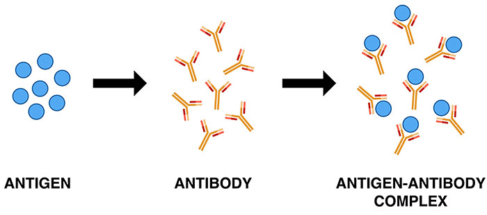
When an antigen and an antibody interact, they form a stable antigen-antibody complex. This binding is one of the immune system's primary methods for protecting itself from pathogens. In addition to being selective, the antigen-antibody interaction has high affinity and cross-reactivity.

Specificity
The spatial and chemical matching of the CDR with the antigen epitope determines antibody-antigen binding, which is sustained by weak non-covalent interactions such as hydrogen bonds, van der Waals forces, electrostatic interactions, and hydrophobic effects. The binding specificity is determined by the precise coupling of the antigen epitope and the antibody variable region sequence.
Affinity
Affinity refers to the strength with which a single antigen epitope binds to the antibody binding site, which has a direct impact on the antibody's neutralizing effectiveness, complement activation efficiency, and signal amplification. High-affinity antibodies sustain stable binding even at low antigen concentrations, which is critical for immunological defense. There is also a concept known as "affinity maturation," achieved through somatic hypermutation in germinal centers, significantly enhancing the antibody's binding efficiency.
The immune system is an arduous network of defense, and antibodies are the "frontline scouts" and "attackers" for discovering and eradicating pathogens and pathological cells while maintaining immune homeostasis. Antibodies' core functions include:
Neutralization of Pathogens
Antibodies specifically target important molecules on the pathogen’s surface (virus envelope proteins, bacterial toxins, and so on), but not host cells. The "blocking" effect doesn’t only obstruct the pathogen’s infected pathway, but also prevents the body from being damaged by the toxin.
Opsonization
Antibodies prevent immune cells (such as neutrophils or macrophages) from acquiring the pathogen when they bind to it. The antibody's Fc component promotes pathogen capture by binding to immune cells' Fc receptors. For instance, antibodies engulf bacterial capsules, opening them up to phagocytes.
Complement Activation
Antibodies (primarily IgG and IgM) attach to the complement system’s C1q molecule through their Fc, triggering the classic complement cascade. This reaction can produce the membrane attack complex (MAC) that directly lyticates the pathogen. The complement cascade also increases inflammation and draws immune cells to the site of infection.
Antibody-Dependent Cellular Cytotoxicity (ADCC)
Following their binding to antigens on the target cell's surface, antibodies attach to NK cells' Fc receptors, triggering the release of granzymes and perforins that kill the target cell. This mechanism is especially important for tumor immunity and viral infections. Therapeutic monoclonal antibodies such as Rituximab employ ADCC to destroy B cell tumors.
Immune Complex Clearance
Antibodies create immune complexes with antigens that are filtered out by phagocytes via complement C3b or Fc receptor interactions. Clearance keeps tissues intact and inflammation at bay.
Mucosal Immunity
Mucosal immunity Secretory IgA (sIgA) is the primary antibody in mucosal immunity, covering the respiratory, digestive, and urogenital systems to form a barrier that hinders pathogen adherence and penetration. IgA can also transfer pathogens away from epithelial cells using a "trap transport" method.
Immune Regulation
Antibodies can tame the immune system, both directly and indirectly, by binding to different receptors and keeping immune activation and tolerance in equilibrium. This role is central to anti-autoimmune health.
Immune Cell Recruitment and Reactivation
The antibodies connect to Fc receptors on immune cells (eosinophils, basophils, and macrophages) via their Fc domain, causing the cells to move to the site of an infection or inflammation. For example, IgE binds to mast cells, causing allergic reactions and regulating the immune system's response to parasites.
Maintenance of Immune Memory
Adaptive immunity relies on antibodies to store memory B cells and long-lived plasma cells (LLPCs) in the body so that it can rapidly attack previously faced pathogens. The long-term pharmacological activity of high-affinity IgG is what underlies long-term vaccine immunity.
The industrial manufacture of antibodies is a highly complex and precise process that includes numerous steps, each of which is crucial to the quality and efficiency of antibody synthesis.
The first step is to select the appropriate antigen and optimize its structure to ensure it can effectively trigger an immune response and specifically bind with the corresponding antibody. This typically includes antigen purification, epitope analysis, and antigen modification to enhance immunogenicity.
The most common method is hybridoma technology, which involves fusing B cells from immunized animals with myeloma cells and selecting hybridoma cells that can secrete specific antibodies. Phage display, cell display, and other techniques are also employed in antibody screening, especially in more efficient and precise screening processes.
Using genetic engineering, the gene for the target antibody can be cloned into an appropriate host cell for expression. Common host cell lines include CHO (Chinese hamster ovary) cells, NS0 cells, and HEK293 cells. Recombinant antibody expression often requires optimizing culture conditions and expression systems to improve yield and antibody quality.
This crucial step aims to isolate the target antibody from the culture medium. Common purification methods include affinity chromatography (using the specific binding between antibody and antigen), ion exchange chromatography, and gel filtration chromatography. During purification, the antibody's structure, function, and purity are rigorously monitored to ensure the final product's quality.
Monoclonal antibodies are highly specific antibodies produced by a single B cell clone. Each monoclonal antibody recognizes and binds to a specific antigen epitope, providing strong targeting capacity. Monoclonal antibodies are employed in numerous applications, including purification, treatments, and diagnostics. They have demonstrated significant potential in antiviral and cancer immunotherapy.
Several B cell lineages produce polyclonal antibodies, which can recognize and bind to several epitopes of the same antigen. Polyclonal antibodies have lesser affinity but are more widely applicable in some experimental conditions (e.g., immunoprecipitation, Western blotting). They are often derived from immunized animals (such as rabbits or goats) and employed when a significant number of antibodies are required.
This technique uses genetic engineering to transplant the Fab region of a mouse monoclonal antibody into the Fc region of a human monoclonal antibody, creating chimeric antibodies. Chimeric monoclonal antibodies have lower immunogenicity and longer half-lives in humans. This technology reduces the immunogenicity of antibodies, enabling better tolerance in humans.
In humanized antibodies, the complementarity-determining regions (CDRs) of a mouse antibody are retained, while the rest of the antibody sequence is replaced with human antibody sequences, minimizing immune reactions caused by the mouse components. Humanized antibodies are suited for long-term therapy and have lower immunogenicity.

By linking cytotoxic drugs to antibodies, ADCs use the specificity of antibodies to deliver drugs precisely to disease sites (such as tumors), improving efficacy and reducing side effects. The antigen targeted by the antibody in ADCs must be highly expressed in tumor tissues and have low or no expression in normal tissues. These antigens are typically classified into tumor-specific antigens (TSAs), which are expressed only in tumor tissues, and tumor-associated antigens (TAAs), which are highly expressed in tumor tissues but low in normal tissues.
Antibodies used in ADCs should meet the criteria of high specificity, strong target binding, low immunogenicity, and low cross-reactivity to ensure efficient uptake of ADCs by tumor cells and to prolong the serum half-life. IgG1 is commonly used in clinical and preclinical ADCs due to its long blood half-life and balanced immune activation. IgG4, which has lower immune activation, is often used for ADC designs requiring high immunogenicity.
Hybridoma technology is the classic and most commonly used method for producing monoclonal antibodies. This process involves fusing myeloma cells with B cells from immunized mice, selecting hybridoma cells that secrete antigen-specific antibodies, and producing and purifying the monoclonal antibodies.
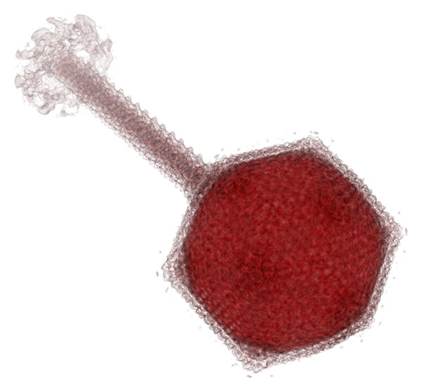
Recombinant DNA technology involves cloning the target antibody gene into a host cell for expression and production. This method provides high flexibility and customization in producing recombinant antibodies.
By displaying antibody fragments on phages or cell surfaces, phage or cell display technologies allow the selection of antibodies with high affinity and specificity, speeding up antibody screening and optimization. Phage display platforms for peptide immunogens are also one of the most cost-effective techniques for discovering peptide vaccines.
The CRISPR-Cas9 system allows precise genetic modifications in host cells, optimizing antibody production or improving antibody function and yield.

Nanobodies (single-domain antibodies) are derived from camelid animals and have smaller molecular weight and stronger stability. They are suitable for therapy and diagnostics and are easier to engineer than traditional antibodies, making them ideal for certain specialized therapeutic needs.
Visualization technologies are used to monitor the antibody production process in real time, such as through fluorescent labeling or mass spectrometry methods, to track the expression, quality, and structural changes during antibody purification.
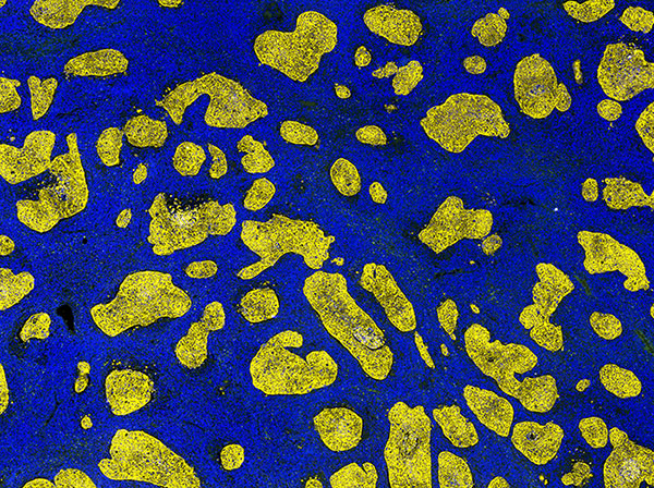
Creative Diagnostics is committed to providing high-quality antibodies with advanced technology and a strict quality control system, ensuring each antibody product exhibits excellent specificity, high affinity, and excellent reproducibility. Our antibodies are tested using ELISA, Western Blot, SPR, and other validation technologies to maintain stable performance under different experimental conditions. We also provide customized services, from antigen design to validation plans, offering targeted antibody solutions to support efficient scientific and industrial applications. Choose Creative Diagnostics for reliable and trustworthy antibody products—visit our Antibody Page to make a purchase today!
Loading ......
