
Inquiry Basket
 Loading ......
The zoonotic pathogen shiga toxin producing E. coli STEC (also known as VTEC) produces shiga toxin (also known as verocytotoxin) which results in fatal human disease that manifests as diarrhoea and haemorrhagic colitis and haemolytic uraemic syndrome (HUS). STEC remains a significant public health concern because these bacteria trigger significant outbreaks and HUS acts as the main reason for acute kidney failure in children. Some certain strains of E. coli produced a cytotoxin which kills vero cells, a continuous cell line derived from the kidney of the African green monkey (Chlorocebus sp). Since it kills vero cell lines, the toxin named as "verocytotoxin" or "verotoxin". Other alternative names are also called such as "shiga-like toxin" or simply "shiga toxin" due to high degree of similarity between verotoxin and the toxins produced by Shigella spp. this terminology lead a synonymous calling as Verocytotoxigenic E. coli (VTEC) or Shigatoxigenic E. coli (VTEC). So Verocytotoxigenic E. coli (VTEC) and Shigatoxigenic E. coli (VTEC) are strains of the bacterium E. coli that produce verocytotoxins also known as Shiga toxins. Not all STEC/VTEC cause illnesses in humans. Only those STEC/VTEC capable of causing severe illnesses in humans are referred to as Enterohemorrhagic E. coli (EHEC). The STEC/VTEC strains that cause haemorrhagic colitis (bloody diarrhoea) belong to the EHEC group of pathogenic E. coli. EHEC pathotype is the most severe pathotype which is capable of leading fatal complications in healthy infected hosts.
The majority of human STEC/VTEC infections stem from serotype O157:H7 according to reports of outbreaks and sporadic cases. The main animal reservoirs of STEC/VTEC exist within ruminant populations where cattle serve as the primary hosts. The primary routes of STEC/VTEC infection occur through contaminated food and water consumption and direct animal contact and human-to-human transmission.
The most common Verocytotoxigenic E. coli (VTEC) is O157:H7 strain. STEC/VTEC are characterized by possession of genes encoding for vtx1 and vtx2, although they carry other virulence genes such as eae and ehxA. Animals are principal reservoirs of STEC/VTEC, and the main route of transmission is fecal-oral. In humans, especially children and elderly, STEC/VTEC cause abdominal cramps associated with diarrhea or dysentery. Complicated cases of STEC/VTEC infection may lead to Hemolytic uremic syndrome (HUS). VTEC serotypes are different than each other with respect to occurrence and symptoms. Serotyping is mainly made using O and H antigens. The outer membrane of an E. coli cell contains lipopolysaccharide (LPS) molecules, that carry; O antigen, a polymer of immunogenic repeating oligosaccharides (1-40 units), Core region of phosphorylated nonrepeating oligosaccharides and Lipid A (endotoxin). The O antigen is used for serotyping E.coli and this O group designations start with O1 and end with O181. Some groups have been removed which are O31, O47, O67, O72, O93 (now K84), O94, and O122; and groups 174 to 181 are provisional (O174 = OX3 and O175 = OX7) or are under investigation (176 to 181 are STEC/VTEC). Moreover, many O groups have subtypes (e.g. O128ab and O128ac). The H antigen is one of the major component of flagella, have role in E. coli movement. It is mainly encoded by the fliC gene. H antigens have 53 identified groups changing between H1 to H56. Serotype Severe disease in humans include O26:H11,O103:H2, O145:NM, O111:NM, O121:H19 and O104:H4. E. coli STEC/VTEC has over 400 serotypes. The serotype O157 is the most frequently isolated serovar from the STEC group. Serovar O157 leads to serious diseases, such as HUS, HC in the humans gastrointestinal tract, due to the production of Stx.
Transmission of STECs to humans occurs through consumption of contaminated foods, such as raw or undercooked ground meat and raw vegetables or direct contact with an infected person. Ruminants, particularly cattle, are the primary reservoirs of STEC worldwide. Moreover, STEC has been isolated from many environments, including drinking water sources. In beef production, the meat production process has been identified as an area where STEC infection can be controlled and prevented. Food handling practices of food supply chain can contribute significantly to STEC transmission. In facilities where meat is processing, contaminated equipment, utensils or surfaces can cross-contaminate cuts of meat, especially ground (minced) beef which mixes trimmings from various parts or sources.
 Figure 1. Multiple routes of VTEC/STEC transmission from different environment sources. (Alhadlaq, 2024)
Gene-encoding virulence factors are similar among STEC serotypes. However, the number/code assigned to individual STEC strains depends on the location of these virulence factors in plasmids, pathogenicity islands (PAIs) and other mobile elements. The PAI LEE possesses several key pathogenic genes, such as the eae gene, which encodes intimin, as well as a type III secretion system and translocated intimin receptor. The gene eae is encoding A/E lesions and located on the LEE, which contributes to STEC pathogenesis by increasing bacterial attachment to intestinal epithelial cells. The LEE and its contents are responsible for A/E lesions induced on host intestinal epithelial cells. The presence of LEE is linked to HUS, at least in some STEC serotypes.
STEC also has two stx genes, stx1 and stx2, that produce the following subtypes: four (Stx1a, Stx1c, Stx1d and Stx1e) and 12 (Stx2a–Stx2l) respectively. Both types of Shiga toxins are AB5 toxins that bind to the glycosphingolipids globotriaosylceramide (Gb3, CD77) and, to a lesser extent, globotetraosylceramide (Gb4), which are found on a variety of human cells, such as glomerular and brain endothelial cells. STEC expresses Stx mainly when the phage becomes lytic during STEC lysis by the host. A combination of stx2a and eae genes is linked to bloody diarrhoea and the development of HUS in infected individuals. STEC pathogenicity is also linked to the presence of haemolysin virulence factors (ehxA, hlyA, e-hlyA and sheA). The ehxA factor is a plasmid-encoded enterohaemolysin and genetically grouped into six subtypes (A–F), which are involved directly in HUS and cases of diarrhoea. It is used as an epidemiological marker to detect STEC serotypes.
The only way to identify all types of VTEC in any kind of test sample is the detection of Verotoxins produced by the bacteria or of the genes associated with VT production. A number of biological (cytotoxicity tests) and serological [ELISA, immunoblot and reversed-passive latex agglutination (RPLA)] tests were developed for identification of Verotoxins produced by the bacteria and genetic methods [polymerase chain reaction (PCR) and DNA-hybridization] are used for identification of VT1 and VT2 (stx1 and stx2) specific DNA sequences. Screening methods, which are not directed to the identification of VTs or their genes (plate indicator media such as SMAC or enterohaemolysin agar, serological typing of putative VTEC isolates) can certainly help in isolation of these pathogens but the suspected VTEC isolates have always to be confirmed as such by identification of VT production itself or by the presence of VT genes.
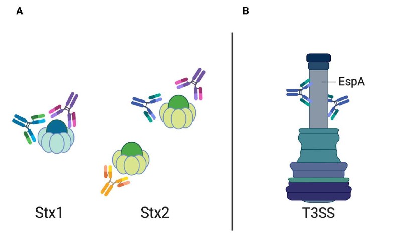 Figure 2. Antibody against STEC-induced diseases. (Mühlen, 2020)
The treatment of STEC infections shows promise through the use of antibody therapy. The administration of Stx-specific antibodies during the early stages of infection leads to Shiga toxin neutralization which protects against disease development. Research with rabbits and bovine colostrum containing Stx1/Stx2 antibodies demonstrated their ability to decrease bacterial colonization while improving survival rates in children with no adverse effects. Scientists propose using transgenic plant-based production of recombinant antibodies as a potential edible therapeutic solution. The therapeutic potential of antibodies extends beyond toxin neutralization because researchers can target bacterial virulence factors. The therapeutic potential of STEC-related diseases becomes more effective when using monoclonal antibodies that block EspA secretion system proteins and camelid antibodies that prevent Tir–intimin interactions to reduce bacterial attachment to intestinal tissue. The combination of these therapeutic approaches demonstrates how antibody-based treatments can effectively combat STEC-related illnesses when administered during the initial stages of infection.
Shiga Toxin 1 vs Shiga Toxin 2
Pathogenic E. coli: Types, Toxins, and Detection Methods
Enteropathogenic E. coli (EPEC)
Enterotoxigenic E. coli (ETEC)
Enterohemorrhagic E. coli (EHEC)
Adherent invasive E. coli (AIEC)
Enteroaggregative E. coli (EAEC)
Diffusely-adhering E. coli (DAEC)
Verocytotoxigenic / Shiga toxin-producing E. coli (VTEC / STEC)
References
| Target | Cat. No. | Product Name | Host | |
| E. coli | DAG-ZL0495 | Inactivated Escherichia coli STEC Culture Fluid | N/A | Inquiry |
| Target | Cat. No. | Product Name | Host | |
| E. coli Verotoxin | DAGB116 | Recombinant E. coli Verotoxin II | E. coli | Inquiry |
| DAGB118 | E. coli Shiga Toxin 2 | E. coli | Inquiry | |
| DAGA-985 | Recombinant E.Coli O157:H7 Shiga Like Toxin-2 Subunit B [His] | E. coli | Inquiry | |
| DAGB117 | E. coli Shiga Toxin 1 | E. coli | Inquiry | |
| DAGA-984 | Recombinant shiga toxin-1 subunit B [His] | E. coli | Inquiry | |
| DAGA-878 | Recombinant shiga toxin 2 subunit b (aa 89,>98%) [His] | E. coli | Inquiry | |
| DAGA-877 | Recombinant shiga toxin 1 subunit b (≥95%) [His] | E. coli | Inquiry |
| Target | Cat. No. | Product Name | Size | Species | Application | Detection Sample | |
| E. coli Verotoxin | DEIA2348 | E. Coli Verotoxin (Fecal) ELISA Kit | 96T | Human | Qualitative | Stool supernatant | Inquiry |
| DEIA1966 | E. Coli Verotoxin 1+2 Ag ELISA Kit | 96T | Qualitative | Stool specimens, broth cultures | Inquiry | ||
| DEIASL162 | Shiga toxins ELISA Kit | 96T | Human | Qualitative | Fecal | Inquiry |
Loading ......
