
Inquiry Basket
 Loading ......
The bacterial pathogen Enteropathogenic Escherichia coli (EPEC) produces severe diarrhea that primarily diarrheal illness by disrupting normal intestinal operations. Scientists discovered EPEC in the mid-20th affects infants and young children in developing nations. The intestinal flora produces EPEC strains which lead to severe century before it became recognized as a major diarrheal pathogen that affects millions of people worldwide.
The medical community established EPEC as the primary cause of infantile diarrhea outbreaks during the 1940s and 1950s in developing nations where it led to fatal summer diarrhea outbreaks. The use of molecular diagnostic tools has enabled scientists to better understand how widespread EPEC infections exist in the population. The combination of enhanced hygiene practices and public health initiatives and breastfeeding promotion has led to a decrease in typical EPEC (tEPEC) infections according to worldwide research. The emergence of atypical EPEC (aEPEC strains has become more widespread throughout the world because they affect both patients with symptoms and people who do not show any symptoms. The World Health Organization reports that EPEC causes between 30-40% of infant diarrhea cases throughout Latin America and Africa and Asia while resulting in high mortality rates in developing countries. The pathogen remains active in daycare centers and pediatric wards even though its occurrence has decreased in developed nations.
Typical EPEC (tEPEC): The E. coli adherence factor (EAF) plasmid with bundle-forming pili (BFP) exists in Typical EPEC (tEPEC) strains which create microcolonies through localized adherence (LA) on intestinal epithelial cells. These strains belong to classical O:H serotypes which primarily lead to acute diarrhea in infants younger than one year.
Atypical EPEC (aEPEC): The EAF plasmid absence in aEPEC strains prevents BFP production while these bacteria exhibit different adherence patterns which include LAL and DA and AA. The genetic makeup of aEPEC strains shows wide diversity because they exist in both diarrheal and asymptomatic people and lead to prolonged diarrhea episodes.
EPEC pathogenesis is complex, involving several virulence factors that facilitate adherence, colonization, and host cell manipulation.
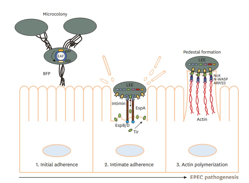 Figure 1. AE lesion formation by Type IV BFP and LEE of EPEC on the small intestine. (Lee, 2022)
The main sign of EPEC infection appears as attaching and effacing (A/E) lesions which develop on intestinal epithelial cells (IECs). The lesions consist of bacterial adhesion to cells while the microvilli disappear and the cytoskeleton creates pedestal structures that support the attached bacteria.
The pathogenicity island known as LEE contains the T3SS apparatus and effectors which create A/E lesions on host cells. The system uses its T3SS to deliver bacterial effector proteins directly into host cells which results in changes to host cell operations.
Exclusive to typical EPEC, BFP facilitates initial bacterial aggregation and microcolony formation on IECs. The plasmid-encoded regulator PerA activates BFP expression. BFP dynamics, regulated by proteins BfpD and BfpF, promote bacterial aggregation and dispersal, essential for spreading infection and intimate adherence via the T3SS.
EPEC also deploys non-LEE encoded effectors (Nle) scattered throughout the genome that modulate host immune responses, protein secretion, and phagocytosis inhibition, enhancing bacterial survival and persistence.
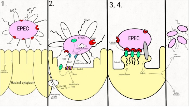 Figure 2. Schematic representation of the EPEC adherence mechanism. (Mare, 2021)
In the first step, EPEC cells express the intimate adhesin intimin, Bfp (bundle-forming pili), and EspA (short filaments surface-associated). Environmental factors regulate the expression of these virulence factors, influencing the site of the bacterial colonization (small/large bowel).
In the second step, EPEC strains adhere to the intestinal epithelium through Bfp and EspA, forming dense microcolonies on the cell's surface, in a pattern described as localized adherence. The type-three-secretion-system creates a pore, enabling the bacteria to inject Tir and a large number (at least 25, up to 50) of effector molecules into the host cell. These effectors facilitate bacterial colonization, immune evasion, and regulate inflammatory response and host cell death. They also activate the host cell-signaling pathways, causing the alteration of the cytoskeleton, leading to the loss of the microvilli. After tyrosine-protein kinase and protein kinase A modifies Tir, it is inserted into the host cell membrane.
Enterocyte effacement and intimate bacterial attachment to the host cell characterizes the third step of EPEC infection. The bacterial cells lose the EspA filaments from their surface. The bacterial intimin (encoded by eae) binds to the modified Tir, causing the intimate attachment of the bacteria to the host cell. In this phase, actin and cytoskeletal elements are accumulated near the site of the bacterial adherence. An autotransporter system, T5SS (type V secretion system), mediates the secretion of EspC, a protein involved not only in epithelial cell cytotoxicity, but also in bacterial replication and biofilm formation.
During the fourth step, the cytoskeletal elements, accumulated near the site of the attachment, leads to the formation of the pedestal structure, characteristic for EPEC. The effector molecules (translocated from the bacteria) disrupt the cell processes, leading, eventually, to cell death.
EPEC infection typically leads to watery, non-inflammatory diarrhea without fever or severe abdominal pain. Atypical strains may cause mild but prolonged diarrhea, particularly in young children. Detection of EPEC relies on molecular techniques targeting virulence genes such as eae and bfpA. Quantitative PCR assays can differentiate between symptomatic and asymptomatic carriage by evaluating bacterial load. However, asymptomatic colonization, especially with aEPEC, complicates diagnosis. There is a pressing need for quick, cost-effective diagnostic tools in endemic regions to facilitate timely treatment and reduce morbidity and mortality among children.
Pathogenic E. coli: Types, Toxins, and Detection Methods
Enteropathogenic E. coli (EPEC)
Enterotoxigenic E. coli (ETEC)
Enterohemorrhagic E. coli (EHEC)
Adherent invasive E. coli (AIEC)
Enteroaggregative E. coli (EAEC)
Diffusely-adhering E. coli (DAEC)
Verocytotoxigenic / Shiga toxin-producing E. coli (VTEC / STEC)
References
| Target | Cat. No. | Product Name | Host | |
| E. coli | DAG-ZL0481 | Inactivated Escherichia coli EPEC 026:K60 Culture Fluid | N/A | Inquiry |
| DAG-ZL0482 | Inactivated Escherichia coli EPEC O 127 : K63 Culture Fluid | N/A | Inquiry | |
| DAG-ZL0483 | Inactivated Escherichia coli EPEC O86:K61 Culture Fluid | N/A | Inquiry |
| Target | Cat. No. | Product Name | Host | Isotype | Application | |
| E. coli Intimin | DPAB-CAB5161 | Anti-E. coli eae/Intimin Polyclonal antibody | E. coli O127:H6 | IgG | ELISA, IHC, WB | Inquiry |
Loading ......
