
Inquiry Basket
 Loading ......
DAEC was the last identified diarrheagenic E. coli pathogroup. It is primarily defined by a characteristic 'diffuse adherence' (DA) pattern observed on HeLa and HEp-2 epithelial cells. The bacteria in this pattern stick to all areas of epithelial cells at equal rates throughout the entire surface. The DAEC pathogroup consists of multiple different E. coli bacterial strains which share no common genetic characteristics. The bacteria require Afa/Dr adhesin genes to establish diarrheal infections in people who do not have underlying health conditions. The Afa/Dr family consists of three adhesins which include Afa and Dr and F1845 and these proteins derive from the afa/dra/daa operons. The infection process of DAEC results in major damage to epithelial cells through Afa/Dr-dependent mechanisms which cause microvilli destruction and enzyme dysfunction of brush border enzymes and disruption of adherens junctions.
 Figure 1. DAEC adhesion to epithelial cells.
(Arikawa et al. 2005 and Cookson and Nataro 1996)
Diffuse Adherence Pattern: DAEC is characterized by its ability to adhere uniformly across the entire surface of epithelial cells, a phenomenon known as diffuse adherence (DA). This distinct pattern is observed on cell lines such as HeLa and HEp-2.
Target Cell Preference: The first step of DAEC pathogenesis occurs when the bacteria attach to intestinal epithelial cells. Research using ex vivo methods demonstrates that DAEC bacteria show superior attachment to M cells which are part of Peyer's patch-associated epithelium compared to the microvilli of Peyer's patch-associated epithelium. The bacteria successfully establish themselves in both human undifferentiated crypt cells and differentiated enterocytes.
Initiation of Pathogenesis: DAEC pathogenesis initiates with its adhesion to intestinal epithelial cells.
Afa/Dr Adhesins and Receptor Binding: Afa/Dr Adhesins and Receptor Binding: The Afa/Dr family of adhesins which includes fimbrial (Dr, F1845) and afimbrial (Afa) types exists on the outer membrane of DAEC bacteria to enable both DA pattern formation on epithelial cells and intestinal epithelial attachment. The adhesins establish connections with particular receptors that exist on host cells.
 Figure 2. General genetic organization of afa/dra/daa operons and assembly of Dr and Afa-III adhesins. (Meza-Segura, 2016)
Induction of Finger-like Projections and Embedding: DAEC attachment to target host cells induces finger-like projections that extend from the surface of infected epithelial cells, effectively embedding the bacteria without complete internalization. This embedding mechanism is believed to provide protection against host-mediated defense mechanisms, antibiotics, and intestinal clearance by peristalsis.
Genetic Organization of Adhesins: The afa/dra/daa operons which produce Afa, Dr and F1845 adhesins consist of five or more genes (A, B, C, D, E). The operon contains five genes which encode a transcriptional regulator and a chaperone and an usher and an invasion protein and an adhesin structural subunit. The D and E subunits exit the cell through the general secretory pathway to reach the periplasm where chaperones in the periplasm help them fold before usher-mediated polymerization.
Secreted Autotransporter Toxin (Sat): The secreted autotransporter toxin (Sat) functions as a major virulence factor. The sat gene appears at higher rates in DAEC strains which cause diarrhea in children compared to DAEC strains from healthy children.
Flagella: Mobile DAEC strains can induce high levels of IL-8 secretion, with purified flagella from Afa/Dr DAEC capable of inducing IL-8 production via TLR5. Since TLR5 is found on the basolateral membrane of polarized epithelial cells, motile DAEC must disrupt tight junctions to allow flagella access to TLR5 and trigger signaling.
Other Virulence Factors: Some DAEC strains carry genes homologous to those of EPEC and EHEC type three-secretion systems, producing EspA, EspB, and EspD homologs, and inducing pedestal formation. Other documented virulence factors include toxins (e.g., astA), siderophores (fyuA, irp2, iuc, iroN), transport systems (shu, modD), and intra-macrophage survival factors (htrA, dsbA).
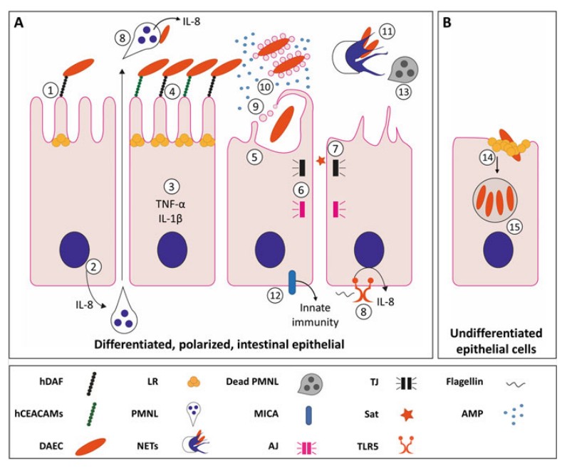 Figure 3. DAEC infection. (Meza-Segura, 2016)
To overcome the limitations of cell adhesion assays, molecular methods that target genes or operon regions of afa and daa are preferred for DAEC identification. These methods ensure more specific detection of DAEC strains. Early DAEC identification relied on DNA probes. PCR Methods: In recent years, PCR methods have become the preferred and more specific approach for DEC identification, including DAEC. Both single and multiplex PCR methods have been developed for this purpose. The first detection methods used diffuse adherence observation but their non-specific nature required scientists to create molecular detection methods. The development of DNA probes brought better results but PCR methods which detect afaC or daaD genes represent the most precise and dependable approach for DAEC identification.
Pathogenic E. coli: Types, Toxins, and Detection Methods
Enteropathogenic E. coli (EPEC)
Enterotoxigenic E. coli (ETEC)
Enterohemorrhagic E. coli (EHEC)
Adherent invasive E. coli (AIEC)
Enteroaggregative E. coli (EAEC)
Diffusely-adhering E. coli (DAEC)
Verocytotoxigenic / Shiga toxin-producing E. coli (VTEC / STEC)
Reference
Loading ......
