
Inquiry Basket
 Loading ......
SARS-CoV-2 and SARS-CoV are 79%-80% homologous at the genomic level. The entire genome is ~30kb in size, and organized into 14 ORFs that code for 29 viral proteins. Two-thirds of the 5' end of the genome encodes for two overlapping polyproteins, pp1a and pp1ab. These are subsequently cleaved by two viral proteases into 16 non-structural proteins (NSPs), responsible for viral replication and transcription. Four ORFs at the 3' end of the genome encode the four structural proteins that were expected: nucleocapsid, spike, membrane and envelope proteins. These proteins mediate virion assembly and also play a role in suppression of the host immune response. A small number of accessory genes that are intermingled between the structural genes encode the accessory proteins. The role of accessory proteins is to modulate viral infection. In contrast to the structural proteins, accessory proteins are not typically part of the virion with the exception of ORF3a and ORF7a.
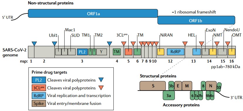 Figure 1. SARS-CoV-2 genome organization, with functional domains shown in rectangles and the prime drug targets emphasized in the outlined box
(Source: Yang H, et al. 2021)
Creative Diagnostics has a wide range of SARS-CoV-2 related antigens, antibodies, and ELISA kits for your diverse detection needs. In addition, our targeted testing products and professional services are also available to help you perform your research/diagnostic tasks with more efficiency and accuracy.
 Figure 2. The life cycle of SARS-CoV-2, including viral entry, replication and transcription, assembly and release. (Source: Yang H, et al. 2021)
The S protein is the principal surface protein of SARS-CoV-2 that binds and fuses with the host cell. It is multi-faceted and its complex structures in multiple conformations mediate viral entry, providing attractive vaccine and antiviral drug design targets.
 Figure 3. Structure of the spike protein trimer.
(Source: Bai C, et al. 2022)
1. Structural Rearrangement and Function
The S protein goes through a large-scale structural transition from a metastable prefusion conformation to a postfusion conformation that mediates fusion between the viral and cellular membranes.
2. Receptor Binding Triggers Fusion
3. Conformational Changes After Binding
4. Formation of the Fusion Core
 Figure 4. A model for membrane fusion induced by the SARS-CoV-2 S protein
(Source: Jackson CB, et al. 2022)
SARS-CoV-2 vaccines from several technology platforms have entered clinical development or received emergency use authorization.
Inactivated whole-virus vaccines


Gene therapy–based vaccines

References
Loading ......
