
Inquiry Basket
 Loading ......
Human cytomegalovirus (HCMV) is a double-stranded DNA virus that belongs to the β-subfamily of herpesviruses. It is the DNA virus with the largest genome among human herpesviruses. The full length of the HCMV genome is approximately 240 kb, consisting of a long unique sequence (UL) and a short unique sequence (US) with inverted repeat sequences at either end. HCMV has a complex proteome and it is estimated that HCMV has more than 200 protein-coding open reading frames. HCMV replication is tightly regulated in multiple steps and gene expression can be divided into immediate early, early and late stages. The HCMV envelope glycoprotein (GP) is the major determinant of tissue affinity and plays an important role in viral entry and cell-to-cell spread.
After the body is initially infected with HCMV, the virus cannot be completely eliminated by the body's immune response and enters a period of latent infection, followed by long-term or intermittent shedding of the virus. HCMV has a variety of latent sites and viral DNA has been detected in monocytes, dendritic cells, megakaryocytes and bone marrow progenitor cells and endothelial cells in the bone marrow. In immunocompromised populations (including organ transplant recipients, haematopoietic stem cell transplant recipients, HIV-infected patients and patients receiving immunomodulatory drugs), latent HCMV infection is more likely to reactivate and invade organs and tissues such as the lungs, gastrointestinal tract, central nervous system and retina, causing serious complications or even death. Cytomegalovirus (CMV) infection can be transmitted by close contact, blood or tissue exposure, perinatal exposure and sexual exposure. The innate immune barrier is the body's first line of defence against CMV entry. If the barrier is damaged or not fully developed (such as the blood-foetal barrier), CMV will enter the body. During the infection process, CMV is first recognised by non-specific immune cells, such as natural killer (NK) cells, monocytes-macrophages and dendritic cells (DCs), initiating an innate immune response. Innate immunity can recognise pathogens and trigger cell signalling cascades, regulate the body's production of interferon (IFN) and pro-inflammatory factors, then trigger the body's antiviral response and activate the adaptive immune response. The adaptive immune response can generate protective immunity against CMV through the direct antiviral effects of specific lymphocytes and the secretion of various cytokines, and control viral replication.
In its long-term coexistence with the host, CMV has evolved a complex anti-immune response mechanism. Not only can it use its huge genome to induce the synthesis of multiple components to participate in the host's anti-infection response, but it can also achieve immune escape by regulating host cells. The immune escape mechanism is an important research topic in the field of CMV. The analysis of its mechanism has important scientific implications for the design of safe and effective new vaccines and the development of antiviral drugs. This article reviews the progress made in the study of the CMV immune escape mechanism.
Interference with Major Histocompatibility Complex
Interference with major histocompatibility complex (MHC) is the major tissue antigen system representing individual specificity, which participates in the regulation of immune response. It can be divided into three categories according to the structure and function of the encoded protein: MHC-I, MHC-II and MHC-Ⅲ.
Interference with MHC-I molecules: The biosynthesis, transport and expression of MHC-I molecules are directly related to the antigen presentation restricted by this type of molecule, thereby affecting the immune response. Studies have confirmed that CMV can downregulate the expression of MHC-I molecules. CMV can also interfere with the synthesis and transport of MHC-I molecules at different links through escape proteins such as US2, US3, US6 and US11, so that they cannot reach the surface of infected cells or express on the cell surface, resulting in CMV-infected cells unable to present endogenous antigens to CD8+ T cells, thereby escaping the killing effect of cytotoxic T cells (CTL). US6 can bind to the antigen processing-related transporter complex to prevent the transport of antigen peptides from the cytoplasm to the endoplasmic reticulum (ER). US3 will hinder the assembly and maturation of MHC-I molecules and their transport from the ER to the cytoplasm. US2 and US11 can bind to the α chain of MHC-I molecules and destroy their structure, or transport newly synthesized α chains from the ER to the cytoplasm through the ubiquitin-dependent pathway, and degrade them under the co-enzymatic hydrolysis of N-glycosidase and proteasome. The phosphorylated protein pp71 encoded by the UL82 gene is packaged in the viral envelope, preventing the assembly of transcriptionally repressive heterochromatin on the viral genome, promoting the transcription and translation of viral genes, and helping immune escape by downregulating the expression of MHC-I molecules and delaying the transport of MHC-I molecules from the ER to the Golgi complex.
Interference with MHC-II molecules: CMV can escape activated CD4+T cells through different pathways. CMV can synthesize a variety of substances to inhibit the intracellular signal transduction required for the expression of MHC-II molecules, such as interleukin-10 (IL-10) and M78 protein. Glycoproteins US2 and US3 can interfere with antigen presentation mediated by MHC-II molecules. US2 binds to MHC-II α chain and assembles MHC-II-α/β/li complex, resulting in the degradation of MHC-II molecules. US3 can compete with the constant chain to bind to MHC-II molecules.
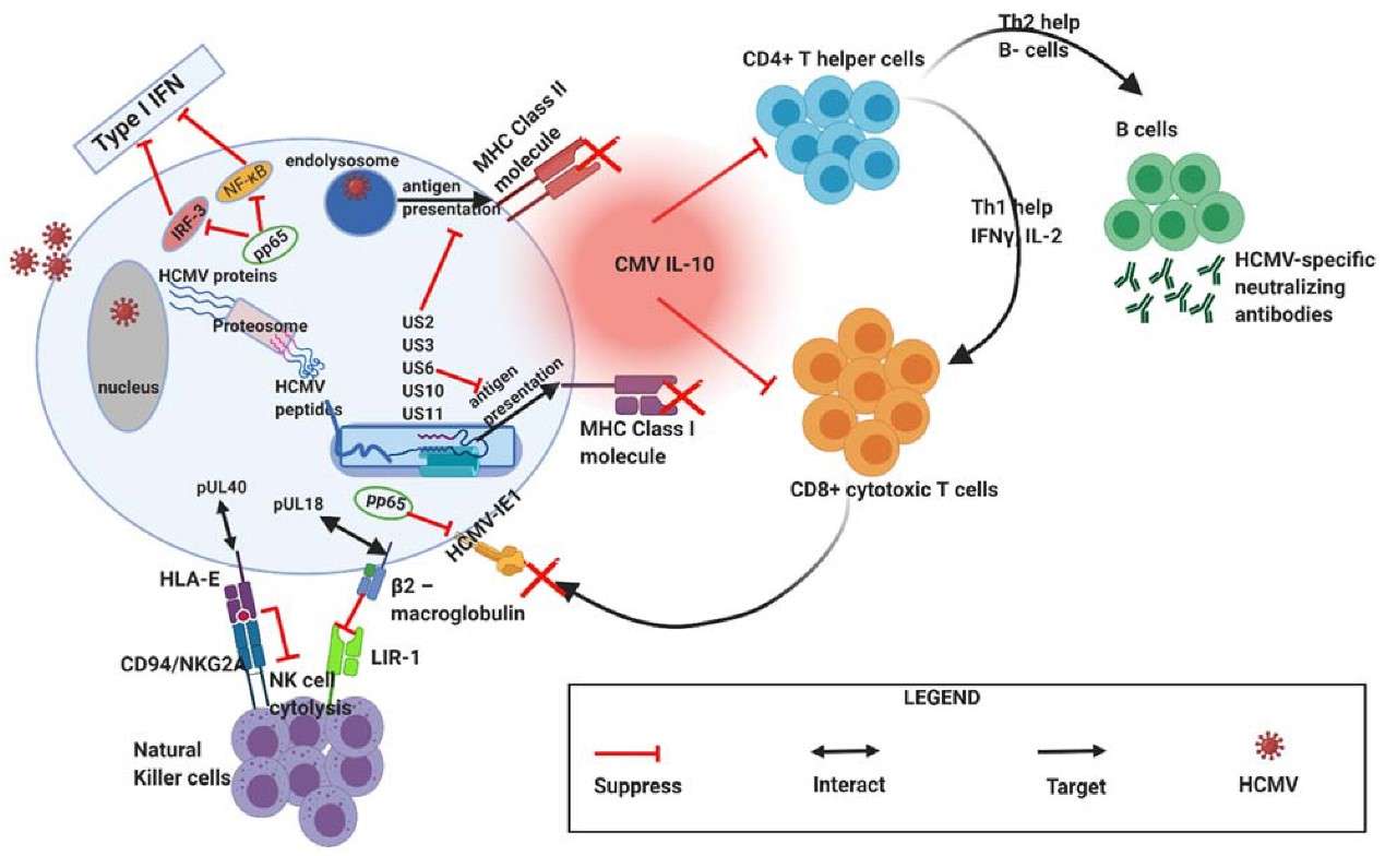 Figure 1. During primary infection, HCMV employs various mechanisms to mediate immune evasion. (Sources: Cox M, et al. 2021)
Due to long-term exposure to persistent antigenic stimulation and inflammation, T cells gradually lose their effector function and begin to lose memory T cell characteristics in chronic infection, a process termed T cell exhaustion. The main manifestations are the gradual loss of effector function (such as cytokine production defects), increased and persistent expression of multiple inhibitory receptors, metabolic dysregulation, and cell transcriptional and epigenetic changes. The increase in soluble immunosuppressive factors and T cell dysfunction are the main causes of T cell exhaustion.
Cytokines: Studies have shown that cytokines and the inflammatory milieu play an important role in the activation and differentiation of T cells. Among these, IL-10 is an important pleiotropic cytokine that has a bidirectional immunoregulatory effect in CMV infection, mainly negatively regulating the virus and helping it to escape the immune system.
Inhibitory receptors: Under normal conditions, PD-1 is generally expressed briefly during effector T-cell activation, after which PD-1 levels return to baseline levels; in chronic infection, PD-1 expression on T cells is maintained at high levels. Studies have shown that PD-1 is expressed at high levels after CMV reactivation in patients with severe sepsis, accompanied by CD8+ T cell exhaustion and a decrease in the proportion of multifunctional CD8+ T cells.
Immunomodulatory or inhibitory cells: CD4+ T cells antagonize CD8+ T cell exhaustion to a certain extent. If the number of CD4+ T cell lymphocytes decreases, the probability of CMV reactivation increases accordingly. Regulatory T cells (Treg) can promote T cell exhaustion through mechanisms such as cell-cell contact, cytokine mediation, and transcription factor regulation. CD8+ T cells are the main target of CMV-specific Treg action. Blocking PD-1 therapy can restore the proliferation ability of CD8+ T cells and reduce T cell apoptosis.
IFN can be divided into three types: type I, type II and type III IFN. Type I IFN, such as IFN-α and IFN-β, can be secreted by most cells; type II IFN includes only IFN-γ, which is mainly secreted by natural killer (NK) cells and T lymphocytes; type III IFN consists of IFN-λ1, IFN-λ2, IFN-λ3 and IFN-λ4. Pattern recognition receptors such as Toll-like receptors, cyclic guanosine monophosphate synthase (cGAS) and interferon stimulator (STING) can recognise pathogen-associated molecular patterns, initiate cell signalling cascades, promote IFN gene activation, and subsequent expression and secretion, thereby exerting antiviral effects. The researchers performed CMV-specific T-cell immune detection in 53 critically ill, non-immunosuppressed patients, and the results showed that IFN-γ was inversely proportional to CMV DNA load. Other researchers used IFN-γ release assays to predict the risk of CMV infection in haematopoietic cell transplant recipients. They followed 241 CMV-seropositive HSCT recipients, all of whom underwent CMV-specific enzyme-linked immunosorbent assays to detect CMV infection within 6 months of transplantation. They found that lower levels of IFN-γ were associated with a higher risk of active CMV infection. Studies have shown that CMV encodes different proteins or uses host cell components to antagonise IFN production and its induced signalling pathways as one of the major strategies for immune escape.
CMV can evade the killing effect of NK cells by regulating the inhibitory or activating signals of NK cells. Inhibitory receptors include CD94/NKG2C in killer cell lectin-like receptors, immunoglobulin-like transcript receptors, etc., while NKG2D and NKp30 are activating receptors. Several genes in the CMV genome, such as UL135, UL142 and UL148, have been shown to inhibit NK cell recognition and killing by different mechanisms. UL135 inhibits the formation of immune synapses by remodelling the actin cytoskeleton; the glycoproteins encoded by UL142 and UL148A inhibit NK cell activation by downregulating the expression of the NKG2D ligand MICA on the cell surface; UL148 affects the expression of the CD2 co-stimulatory molecule CD58 (LFA-3). In addition, m02 and m145 family members in CMV can also express MHC-I molecular analogs, bind to NK cell inhibitory receptors, inhibit NK cell activation and protect infected cells from being killed; UL40 can increase host HLA-E expression, bind to NK cell inhibitory receptor NKG2A, inhibit NK cell activation, and UL40 variants can alter HLA-E binding peptides and stimulate proliferation of NKG2C+NK cells. At present, research into the mechanism of NK cell immune escape is mostly focused on individual genes and products, and the interaction between different ULs and variants and their effects on NK cells are still unclear.
DC are the major antigen-presenting cells with abundant MHC-I, MHC-II, co-stimulatory molecules (CD80, CD86 and CD40, etc.), intercellular adhesion molecules and lymphocyte function-related molecules on their membrane surface, stimulating the activation and proliferation of initial T cells and playing an important role in both initial and sustained immune responses. Currently, DCs present in lymphoid tissues, blood and other non-lymphoid tissues are collectively referred to as classical DCs (cDCs); DCs capable of secreting high levels of IFN-Ⅰ are referred to as plasmacytoid DCs (pDCs), which have different phenotypes and functions. Studies have confirmed that DCs have a dual role in the CMV infection process in mice. While generating an immune response, they specifically downregulate the role of antigen presentation on the surface proteins of initial T cells and initiate immunosuppression. cmvIL-10 and LAcmvIL-10 are IL-10 homologs encoded by CMV UL111A that can mediate DC immune escape through a variety of mechanisms. cmvIL-10 can not only inhibit the expression of MHC-I and class II molecules and co-stimulatory molecules on the surface of DCs, but also inhibit the activation of anti-apoptotic genes (bcl-1, bcl-2 and bcl-x), leading to rapid apoptosis of DCs; LAcmvIL-10 has some functions of cmvIL-10, such as inhibiting the expression of MHC-II molecules on the surface of DCs. In addition, inflammatory inhibitory cytokines such as IL-10, TGF-β, Toll-like receptor 2 (TLR2) and TLR7 can trigger the molecular microenvironment leading to low expression of CD80, CD86 and MHC-II, thereby transforming immature DC into tolerised dendritic cells (tolDC). tolDC will inhibit the expression of some transcriptional molecules such as immunoglobulin-like transcripts, thus playing a key role in establishing and maintaining tolerance.
CMV can directly interfere with viral antibody production and effector functions. Studies have shown that the glycoproteins gp34 and gp68 encoded by the CMV RL11 and UL118-UL119 genes can competitively bind to the FcγR on immune cells at the IgGC terminus, blocking the binding of the viral protein-IgG complex to receptors on the surface of phagocytes and NK cells. By blocking antibody-dependent cell-mediated cytotoxicity and the classical complement activation pathway, removal of these two genes significantly enhances the ability of anti-CMV polyclonal IgG antibodies to activate FcγR and increase the ability of immune cells to clear the virus.
References
| Target | Cat. No. | Product Name | Size | Species Reactivity | Application | Detection Sample | |
| CMV | DEIA-S21CM | CMV IgM ELISA Kit | 96T | Human | Qualitative | serum | Inquiry |
| DEIA013 | Cytomegalovirus IgG ELISA Kit | 96T | Human | Qualitative | serum | Inquiry | |
| DEIA326 | Cytomegalovirus IgG ELISA Kit | 96T | Quantitative | serum, plasma | Inquiry | ||
| DEIA462 | Human CMV IgA ELISA Kit | 96T | Human | Quantitative | Serum | Inquiry | |
| DEIA463 | Human CMV IgG ELISA Kit | 96T | Human | Semi-quantitative | immunocompromised patients | Inquiry | |
| DEIA464 | Human CMV IgG(CSF) ELISA Kit | 96T | Human | Semi-quantitative | Serum | Inquiry | |
| DEIA466 | Cytomegalovirus (CMV) IgM ELISA | 96T | Human | Semi-quantitative | Human serum or EDTA, heparin or citrate plasma | Inquiry | |
| DEIA401 | CMV IgG ELISA Kit | 96T | Human | Semi-quantitative, Qualitative | serum, plasma | Inquiry | |
| DEIA402 | Human CMV-IgM(cytomegalovirus-Immunoglobulin M) ELISA Kit | 96T | Human | Qualitative | Serum, plasma | Inquiry | |
| DEIA2274 | Human CMV IgM ELISA Kit | 96T | Human | Qualitative | serum | Inquiry | |
| DEIA2275 | Human CMV IgG ELISA Kit | 96T | Human | Quantitative | Serum | Inquiry | |
| DEIA465 | Human CMV IgG and IgG avidity ELISA kit | 96T | Human | Qualitative | Serum, plasma | Inquiry | |
| DEIA326R | Human CMV IgG ELISA RUO | 96T | Human | Quantitative and qualitative | serum and plasma (EDTA, lithium heparin or citrate plasma) | Inquiry | |
| DEIA2275S | Cytomegalovirus IgG ELISA kit | 96T | Human | Quantitative and Qualitative | Serum, Plasma and Cell Culture Supernatants | Inquiry | |
| DEIA-NS2307-106 | Human CMV-IgG(cytomegalovirus-Immunoglobulin G) ELISA Kit | 96T | Human | Quantitative | serum, plasma, tissue homogenates and other biological fluids. | Inquiry | |
| DST-H018 | (CMV) Cytomegalovirus IgM/IgG Antibody Rapid Test | 96T | Human | Qualitative | Serum, plasma | Inquiry | |
| DEIAPV4 | Cucumber Mosaic Virus (CMV) ELISA Kit | 500T/1000T/5000T | Qualitative | host plans | Inquiry |
Loading ......
