
Inquiry Basket
 Loading ......
Protein oxidation is a chemical process that involves the modification of proteins by reactive oxygen species (ROS) or reactive nitrogen species (RNS), collectively referred to as oxidative species. ROS and RNS are generated naturally in cells as byproducts of normal metabolism or can be produced in response to external factors such as environmental stress, radiation, or inflammation. While low levels of oxidative species are involved in normal cellular signaling and regulation, excessive production or impaired antioxidant defense mechanisms can lead to oxidative stress and damage to cellular components, including proteins.
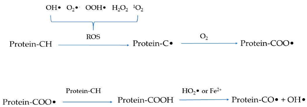 Figure 1. Schematic representation of the mechanism of protein oxidation.
(Source: Domínguez, R. et al., 2021)
Proteins are composed of amino acids, and oxidation primarily affects amino acid residues within the protein structure. The most susceptible amino acids to oxidation include cysteine, methionine, histidine, tyrosine, and tryptophan. These modifications may disrupt the structural integrity of proteins, impair their biological activity, or lead to aggregation and misfolding. Such disruptions can contribute to the development and progression of various diseases, including neurodegenerative disorders, cardiovascular diseases, cancer, and age-related conditions.
Protein oxidation can occur through various mechanisms, each resulting in distinct oxidative modifications and consequences for protein structure and function. Understanding the different protein oxidation mechanisms is crucial for comprehending the diverse molecular events associated with oxidative damage. Some key protein oxidation mechanisms include:
The oxidation of sulfur-containing amino acids, such as cysteine and methionine, is a prominent protein oxidation mechanism. Cysteine residues contain highly reactive thiol (-SH) groups that are particularly susceptible to oxidation. Oxidation of cysteine can result in the formation of disulfide bonds (S-S) or the generation of sulfenic acid (-SOH), sulfinic acid (-SO2H), or sulfonic acid (-SO3H) derivatives. These modifications can disrupt protein structure, alter protein-protein interactions, and impact enzymatic activities. Methionine residues can be oxidized to methionine sulfoxide, leading to changes in protein conformation and function.
Aromatic amino acids, such as tyrosine and phenylalanine, are susceptible to oxidative modifications. Tyrosine residues can be nitrated or chlorinated, resulting in the formation of 3-nitrotyrosine or 3-chlorotyrosine, respectively. These modifications can affect protein signaling, enzymatic activity, and protein-protein interactions. Phenylalanine residues can undergo hydroxylation, leading to the formation of 3-hydroxyphenylalanine.
Lipoxidation involves the reaction of proteins with lipid peroxidation products, which are generated during the oxidative degradation of lipids. Lipid peroxidation products, such as 4-hydroxynonenal (HNE) and malondialdehyde (MDA), can react with proteins, leading to the formation of covalent adducts. Lipoxidation can impair protein function, induce protein aggregation, and contribute to the development of various diseases, including cardiovascular diseases and neurodegenerative disorders.
Glycoxidation refers to the oxidation of proteins by reactive aldehydes derived from the oxidation of sugars, particularly glucose. This process leads to the formation of advanced glycation end products (AGEs), which can cross-link proteins and alter their structure and function. Glycoxidation plays a significant role in age-related diseases, such as diabetes and neurodegenerative disorders.
Protein carbonylation is a common oxidative modification that occurs when proteins are exposed to ROS or reactive carbonyl species. Carbonylation involves the formation of carbonyl groups (C=O) in amino acid residues, such as lysine, arginine, proline, and threonine. Carbonylated proteins are often associated with oxidative stress and aging. The measurement of protein carbonylation levels is frequently used as a surrogate marker for overall protein oxidation.
Protein oxidation occurs due to the action of reactive oxygen species (ROS). After oxidation, molecular changes in proteins can be studied at the level of individual proteins, peptides, or modified amino acids. To analyze these changes, different techniques are available depending on the specific analytes of interest. Among these methods, mass spectrometry stands out as the most accurate technique. It enables precise analysis and should be increasingly utilized for studying protein oxidation in biological samples.
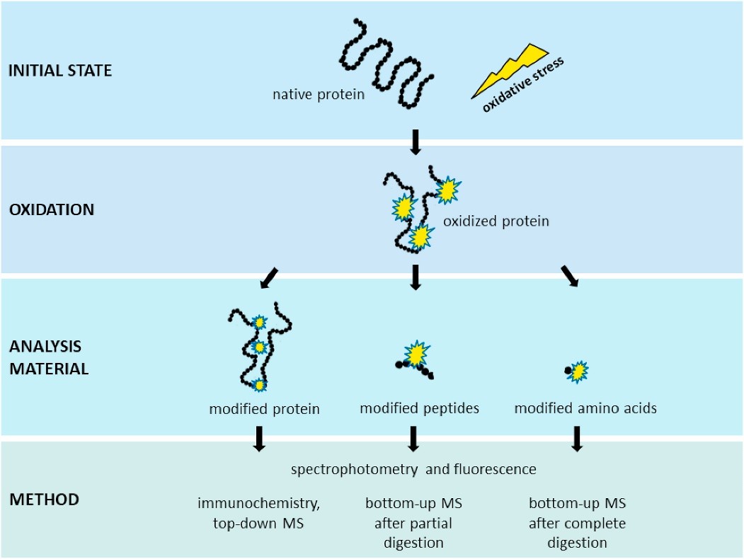 Figure 2. Analysis of oxidative protein modifications.
(Source: Kehm, R. et al., 2021)
Analyzing specific oxidation products is crucial for uncovering the chemistry of oxidation and providing information about the proteins involved. Different analyses can offer quantitative or semi-quantitative data depending on the desired approach. These techniques include:
Protein oxidation has been extensively studied in relation to various human diseases, including diabetes, cardiovascular disease (CVD), cancer, atherosclerosis, arthritis, and neurodegenerative diseases. These conditions are known to increase with age, suggesting a simultaneous occurrence of protein oxidation and aging-related diseases. Therefore, the identification of reliable biomarkers of protein oxidation is crucial for understanding disease mechanisms, assessing disease status, and developing potential treatments. Biomarkers used in clinical applications must meet specific requirements such as sensitivity, specificity, reproducibility, and reliability. The most commonly used and chemically stable biomarkers of protein oxidation in routine clinical diagnostics include:
Studying biomarkers of protein oxidation in the context of human disease and aging has several implications:
References
For research use only, not for use in diagnostic procedures.
| Target | Cat. No. | Product Name | Size | Species Reactivity | Application | Detection Sample | |
| Homocysteine | DEIABL3 | Homocysteine ELISA kit | 96T | Human | Quantitative | plasma, serum | Inquiry |
| DEIA1724 | Human HCY(Homocysteine) ELISA Kit | 96T | Human | Quantitative | serum, plasma, tissue homogenates, other biological fluids | Inquiry | |
| DEIA3576 | HCY(Homocysteine) ELISA Kit | 96T | Universal | Quantitative | Serum, plasma, tissue homogenates and other biological fluids. | Inquiry | |
| DEIABL3-2 | Homocysteic acid ELISA Kit | 96T | Quantitative | Serum, plasma, tissue homogenates, urine | Inquiry | ||
| Tyrosine | DEIANS071 | Nitrotyrosine ELISA Kit | 96T | N/A | Quantitative | cell lysates, serum, plasma and purified proteins | Inquiry |
| DEIA3981 | Nitrotyrosine ELISA Kit | 96T | Human | Quantitative | Cell Lysate, Other biological fluids, Plasma, Serum, Urine | Inquiry | |
| DEIA087J | Nitrotyrosine ELISA Kit | 96T | Human | Quantitative | EDTA plasma, serum | Inquiry | |
| DEIA088J | Nitrotyrosine ELISA Kit | 96T | Human | Quantitative | stool, serum, EDTA plasma | Inquiry | |
| DEIA100J | Tyrosine ELISA Kit | 96T | Human | Quantitative | EDTA plasma, serum | Inquiry | |
| DEIA-PYR064 | Tyrosine Colorimetric Assay Kit | 100T | Quantitative | serum, plasma, urine, other body fluids | Inquiry | ||
| Tryptophan | DEIA05750 | Tryptophan ELISA Kit | 96T | Quantitative | urine, plasma, serum | Inquiry | |
| DEIA074J | Tryptophan ELISA Kit | 96T | Quantitative | cell culture media | Inquiry | ||
| Phenylalanine | DEIA-XY8 | Phenylalanine neonate (with Membrane Plate) ELISA kit | 192T | Quantitative | blood | Inquiry | |
| DEIA-XY11 | Phenylalanine neonate (without Membrane Plate) ELISA kit | 192T | Quantitative | blood | Inquiry | ||
| DEIA098J | Human Phenylalanine ELISA Kit | 96T | Human | Quantitative | EDTA plasma and serum | Inquiry | |
| 4-HNE | DEIA-BJ2157 | Rat 4 Hydroxynonenal ELISA Kit | 96T | Quantitative | Serum, plasma, cell culture supernatants, body fluid and tissue homogenate | Inquiry | |
| DEIA-BJ2812 | Porcine 4 Hydroxynonenal ELISA Kit | 96T | Quantitative | Serum, plasma, cell culture supernatants, body fluid and tissue homogenate | Inquiry | ||
| DEIA-BJ2486 | Mouse 4-HNE(4-Hydroxynonenal) ELISA Kit | 96T | Mouse | Quantitative | serum, plasma, cell culture supernatant and other biological samples. | Inquiry | |
| DEIA-BJ323 | 4-HNE(4-Hydroxynonenal) ELISA Kit | 96T | Universal | Quantitative | Serum, plasma, tissue homogenates, other biological fluids | Inquiry | |
| DEIA-NS2307-31 | Human 4-HNE(4-Hydroxynonenal) ELISA Kit | 96T | Human | Quantitative | serum, plasma, tissue homogenates and other biological fluids. | Inquiry | |
| MDA | DEIASL349 | Rat Malondialdehyde ELISA Kit | 96T | Quantitative | serum, plasma, tissue homogenates and other biological fluids | Inquiry | |
| DEIA3918 | MDA(Malondialdehyde) ELISA Kit | 96T | Universal | Quantitative | Serum, plasma, tissue homogenates and other biological fluids. | Inquiry | |
| CML | DEIA-BJ2094 | Rat Carboxymethyl lysine ELISA Kit | 96T | Quantitative | Serum, plasma, cell culture supernatants, body fluid and tissue homogenate | Inquiry | |
| DEIA-BJ2409 | Mouse Carboxymethyl lysine ELISA Kit | 96T | Quantitative | Serum, plasma, cell culture supernatants, body fluid and tissue homogenate | Inquiry | ||
| DEIA-BJ2780 | Porcine Carboxymethyl lysine ELISA Kit | 96T | Quantitative | Serum, plasma, cell culture supernatants, body fluid and tissue homogenate | Inquiry | ||
| DEIA-XYZ1 | Human CML(Carboxymethyl Lysine) ELISA Kit | 96T | Human | Quantitative | Serum, plasma, tissue homogenates and other biological fluids. | Inquiry | |
| DEIABL517 | Anti-CML Mouse autoAntibody ELISA Kit | 96T | Quantitative | serum, plasma | Inquiry | ||
| DEIABL518 | Anti-CML Human autoAntibody ELISA Kit | 96T | Semi-Quantitative | serum, plasma | Inquiry | ||
| DEIABL519 | Anti-CML Rat autoAntibody ELISA Kit | 96T | Quantitative | serum, plasma | Inquiry |
Loading ......
