
Inquiry Basket
 Loading ......
Neural markers play a crucial role in neuroscience research, allowing scientists to identify and study different cell types within the nervous system. Understanding the specific markers expressed by neurons, neural stem cells, and glial cells is essential for unraveling the complexities of the brain and advancing our knowledge of neurological disorders.
At present, the field of neurobiology is in a stage of rapid development. Antibodies, proteins, ELISA kits, etc., as important research tools, play an indelible role in nerve cell markers and neurological disease signaling pathways. Creative Diagnostics assists in the research of neural-related markers and provides customers with a wide range of high-quality products, services, and professional technical guidance.
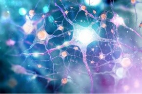
Neurons are the basic structural and functional units of the nervous system. Their main parts include dendrites, cell body, axon, and cell membrane. Dendrites are shaped like many-branched branches, interspersed with many dendrites, and thus have the potential to receive input from many other cells. There is a nucleus in the cell body, and most of the organelles that maintain cell life are in it. The axon is the output end of the cell, extending from the cell body, and is generally very long. Many axons are wrapped in myelin, which acts to insulate them from the flow of information from other cells.
According to the different neurotransmitters released by neurons, neurons are divided into cholinergic neurons, dopaminergic neurons, GABAergic neurons, glutamatergic neurons, and serotonergic neurons.
Efferent nerves that can synthesize acetylcholine and release acetylcholine at the end when excited are called cholinergic nerves. Cholinergic neurons in the cortex and other brain regions are involved in the regulation of sleep, motor function, aggression, and especially higher functions such as learning and memory.
Related targets: choline acetyltransferase, acetylcholinesterase, and vesicular acetylcholine transporter.
Dopaminergic neurons are neurons that contain and release dopamine as a neurotransmitter. Experimental data show that dopaminergic neurons are found in different brain regions in mammals. The highest levels are found in the caudate nucleus and the nucleus accumbens, followed by the substantia nigra and globus pallidum.
Related targets: tyrosine hydroxylase, dopamine transporter, FOXA2, GIRK2, Nurr1, LMX1B.
GABA is the abbreviation of γ-aminobutyric acid, which is an important neurotransmitter in the central nervous system. GABAergic neurons refer to the part of nerve cells that mainly use GABA as a transmitter. GABA is an inhibitory neurotransmitter that has effects on learning and memory as well as sleep.
Related targets: GABA transporter 1, GABAB receptors 1 and 2, GAD65, and GAD67.
Glutamatergic neurons are the most important excitatory neurons in the central nervous system. When glutamate is transported by vesicular glutamate transporters (VGLUTs) into synaptic vesicles, which then fuse with the presynaptic membrane, glutamate is released into the synaptic gap and acts as a neurotransmitter.
Related targets: VGLUT1, VGLUT2, NMDAR1, NMDAR2B, glutaminase, and glutamine synthetase.
5-hydroxytryptamine was first discovered in serum, also known as serotonin, widely present in mammalian tissues, especially in the cerebral cortex and synapses, and it is also an inhibitory neurotransmitter. In peripheral tissues, serotonin is a strong vasoconstrictor and smooth muscle contraction stimulator. In the body, 5-hydroxytryptamine can be catalyzed by monoamine oxidase into 5-hydroxychromaldehyde and 5-hydroxyindoleacetic acid and then excreted with urine.
Related targets: tryptophan hydroxylase, serotonin transporter, and Pet1.
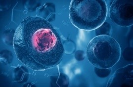
Neural stem cells are a kind of mother cells with division potential and self-renewal ability and have the potential to differentiate into neurons, astrocytes, and oligodendrocytes so that they can produce a large number of brain cell tissues and perform self-renewal. Neural stem cells are sufficient to provide a large population of brain tissue cells.
Related targets: Nestin, SOX2, E-cadherin/CDH1, HES1, HES3, NOTCH1, Occludin/OCLN.
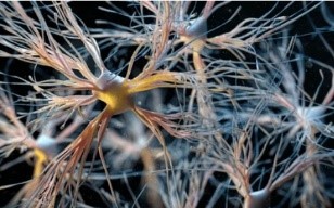
Glial cells often referred to as the "support cells" of the nervous system, play essential roles in maintaining neuronal health and function. They provide structural support, regulate the extracellular environment, and contribute to processes such as myelination and immune response. Distinguishing between different glial cell types relies on specific markers that highlight their distinct functions. There are three types of glial cells.
Astrocytes are the most widely distributed type of cells in the mammalian brain, and they are also the largest type of glial cells. These cells are star-shaped, with many long and branched processes emanating from the cell body, stretching and filling between the cell body of the nerve cell and its processes, supporting and separating the nerve cells. There are two types of astrocytes: protoplasmic and fibrous.
Related targets: GFAP, EAAT2/GLT-1, EAAT1/GLAST, S100 beta, glutamine synthetase, ALDH1L1.
Microglia is a type of mononuclear macrophage resident in the central nervous system (CNS). It is small and short rod-shaped, with several rod-like protrusions, and the surface of the protrusions is rough. Microglia are widely distributed throughout the brain and spinal cord, accounting for 5-20% of all glial cell numbers in the CNS. The normal number and function of microglia are important for maintaining the homeostasis of the nervous system.
Related targets: TMEM119, CD45, CD11b, CX3CR1, IBA1, F4/80, CD68, CD40.
Oligodendrocytes are smaller than astrocytes, with short and few processes and round, small and dense nuclei. Under the electron microscope, the cytoplasm has a high electron density and mainly contains mitochondria, nucleosomes, and microtubules. The main function of oligodendrocytes is to surround axons in the CNS, form an insulating myelin sheath structure, assist in the rapid and efficient transmission of bioelectrical signals, and maintain and protect the normal function of neurons.
Related targets: OLIG2, A2B5, NG2/MCSP/CSPG4, Oligodendrocyte Marker O4, PDGFRA, APC, and Nogo-A/RTN4.
Loading ......
