
Inquiry Basket
 Loading ......
Alzheimer's disease (AD) is a progressive neurologic condition that leads to the decline of cognitive functioning and eventual death. There is currently no cure. Proposed causes of AD include the amyloid hypothesis, which suggests that it is caused by a buildup of amyloid-beta and tau proteins in the brain, leading to cell death. Recent diagnostic tools focus on amyloid and tau proteins as potential markers of the disease, and new treatments are also focusing on amyloid and tau formation. Earlier diagnosis of AD allows time for planning for care and support needs before symptoms worsen. It also allows for both drug and non-drug treatments to be used earlier, which may prolong time with a higher quality of life.
There are numerous proposed and tested methods of diagnosis for AD in the early stages of the disease. These include questionnaires, imaging, CSF biomarkers, blood testing, olfactory biomarkers, salivary testing, and retinal and ocular testing. Some methods of diagnosis are in current use, while others are still in development.
Diagnosis of Cognitive Impairment
There are cognitive tests used for the diagnosis of AD, such as the General Practitioner Assessment of Cognition (GPCOG), the Memory Impairment Screen (MIS), the Mini-Cog test, the Mini-Mental State Examination (MMSE) for dementia, the Clinical Dementia Rating scale-Sum of Boxes (CDR-SB), and the Alzheimer's Disease Assessment Scale-Cognitive Subscale (ADAS-Cog). These cognitive tests are assessed by the patient's attending physician, who only considers functional changes caused by intellectual impairment, ignoring other factors such as physical illness, depression or personality changes, and is somewhat subjective. In addition, these tests are only used to monitor cognitive changes after diagnosis and determine the therapeutic effect, and cannot diagnose early Alzheimer's disease in people without any symptoms.
Brain imaging/Neuroimaging
Neuroimaging is regularly used today for early detection of Alzheimer's. The main imaging auxiliary examination methods for AD include medical imaging technologies such as computed tomography (CT), magnetic resonance imaging (MRI) and positron emission tomography (PET-CT). On the one hand, structural imaging examinations can help rule out other brain diseases or dementia subtypes such as meningioma and subdural hemorrhage. On the other hand, they can be used to predict the transition from mild cognitive impairment to Alzheimer's disease. Functional imaging examinations such as Aβ-PET and tau-PET can show the distribution of marker substances in the brain, thereby assisting in judging and assessing the risk and course of AD.
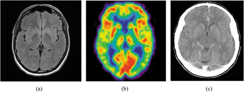 Fig. 1 Examples of brain images: a) MRI b) PET scan c) CT scan
Structural imaging Structural imaging provides information about the shape, position or volume of brain tissue. Structural techniques include MRI and CT. Having shown that the brains of people with Alzheimer's shrink significantly as the disease progresses, structural imaging research also has shown that shrinkage in specific brain regions such as the hippocampus may be an early sign of Alzheimer's. Scientists now have some agreement on standardized values for brain volume loss (sometimes measured over time) that would indicate the presence of disease or disease progression.
 Fig. 2 Representative MRI scans in different types of dementia.
Functional imaging Functional imaging reveals how well cells in various brain regions are working by showing how actively the cells use sugar or oxygen. Functional techniques include PET and functional MRI (fMRI). Functional imaging research suggests that those with Alzheimer's typically have reduced brain cell activity in certain regions. For example, studies with fluorodeoxyglucose (FDG)-positron emission tomography (PET) indicate that Alzheimer's is often associated with reduced use of glucose (sugar) in brain areas important in memory, learning and problem-solving. According to Medicare recommendations, an FDG-PET scan is considered a reasonable test for people with a recent diagnosis of dementia and documented cognitive decline of at least six months who meet diagnostic criteria for both Alzheimer's and frontotemporal dementia.
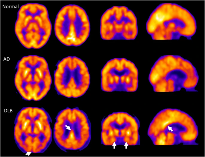 Fig. 3 18F-Fluorodeoxyglucose (FDG) PET in Alzheimer's disease (AD) and Dementia with Lewy Bodies (DLB).
Molecular imaging Molecular imaging uses highly targeted radiotracers to detect cellular or chemical changes linked to specific diseases. Molecular imaging technologies include PET and fMRI. Molecular imaging is among the most active areas of research aimed at finding new approaches to diagnose Alzheimer's in its earliest stages. Molecular strategies may detect biological clues indicating Alzheimer's is under way before the disease changes the brain's structure or function, or takes an irreversible toll on memory, thinking and reasoning. Molecular imaging also may offer a new strategy to monitor disease progression and assess the effectiveness of next-generation, disease-modifying treatments. Several molecular imaging compounds are being studied, and four have been approved for clinical use:
Florbetaben (Neuraceq®), Florbetapir (Amyvid®) and Flutemetamol (Vizamyl®) have been approved for detection of beta-amyloid in the brain.
Flortaucipir F18 (Tauvid®) has been approved for detection of tau in the brain.
Cerebrospinal fluid (CSF) tests
CSF is a clear fluid that bathes and it may be more useful than other biomarkers such as those found via blood tests because of its connection to the brain and relative separation from the rest of the body. Similar to PET imaging, CSF biomarkers include amyloid-beta deposition (amyloid-beta1–42), tau tangle formation (P-tau181), and loss of neuronal function (T-tau). In the CSF, amyloid-beta1–42 decreases throughout the course of the disease, and T-tau and P-tau181 increase. If all these biomarkers are within the normal reference range, it can help rule out AD. Another potential marker is neurofilament light (NfL), an increased level of which has been found in neurodegenerative diseases such as Alzheimer's.
CSF testing for AD-related biomarkers is promising because of its high sensitivity and specificity compared with healthy controls or to autopsied people. CSF testing is not routinely recommended for people who do not have symptoms of cognitive decline because a small percentage of people 85 years and older who are cognitively healthy can have similar testing results as people with AD.
Blood tests
Blood and plasma biomarker tests have been explored as an alternative to the more invasive CSF biomarker tests. Blood tests may also allow for potential tracking of disease biomarkers because repeated collection is relatively easy compared with other methods of biomarker collection. Potential biomarkers include upstream and early biomarkers such as: plasma amyloid-beta species (e.g., amyloid-beta peptides, ratio of amyloid-beta peptides, amyloid-beta oligomers), plasma tau forms (e.g., total tau proteins, phosphorylated tau protein). And downstream biomarkers, such as: neurodegeneration biomarkers (e.g., plasma neurofilament light, plasma synaptic proteins), neuroinflammation biomarkers (e.g., cytokines, chemokines), exosome biomarkers (e.g., microRNAs in exosomes, insulin resistance-related biomarkers in exosomes, lysosomal proteins in exosomes, other molecules in exosomes), genomic and molecular analysis biomarkers (e.g., sphingolipids, acylcarnitine, and glycerophospholipids of lipids), metal-based biomarkers (e.g., copper and ceruloplasmin, iron and ferritin, aluminum, manganese, and zinc). Other options include genetic biomarkers (e.g., familial genes such as the APP gene, PSEN1 gene, and PSEN2 gene) or combinations of these.
The currently available tests may predict the presence of amyloid changes in the brain or the presence of neurodegenerative disease or neuronal damage. These blood tests cannot be used as a stand-alone test to diagnose AD or any other dementia; they will be used as part of a diagnostic workup with other exams.
Salivary tests
Saliva shares 40% of the diagnostic protein biomarkers with blood but gathering saliva samples is less invasive and often cheaper. Relevant biomarkers include salivary acetylcholinesterase, salivary amylase-beta42, salivary tau, salivary trehalose and lactoferrin, salivary metabolites, and the salivary microbiome (i.e., bacteria and microbes in the mouth). Salivary amylase-beta42 was the most reliable biomarker, but this research area is still relatively new. More research is required to confirm the usefulness of salivary biomarkers for AD.
Retinal and Ocular tests
The eye is an extension of the central nervous system (a "window to the brain") which is another focus for diagnosis of AD. Eye diseases and AD also share many risk factors. Deposition of amyloid on the retina of the eye is of particular interest to researchers looking into the early identification of AD. Using optical coherence tomography (a noninvasive test that takes photographic cross-sections of the retina), a clinician can look at the layers of cells in the retina and compare the thickness of the cell layer with control groups. Individuals with AD often have thinner retinal nerve fibre layers. One of the challenges with retinal changes for the identification of AD is creating a consistent test for detection of the disease. Retinal thinning also occurs in other eye diseases, such as glaucoma and diabetic retinopathy, and distinct changes related to AD have not been identified or confirmed.
Genetic risk profiling
Genetic testing for AD is used to determine the potential risk of developing AD, but it is not a biomarker for diagnosis. The genes that influence risk of development of AD are called "susceptibility genes", and include APOE4, and the other familial genes noted previously. Genetic testing for APOE-e4, the strongest risk gene in some populations, is included in some clinical trials to identify participants at high risk for Alzheimer's disease or risk side effects that may be associated with approved treatments.
As more effective treatments are developed, genetic profiling may become a valuable risk assessment tool for wider use.
| Aβ42 | ||||
| Target | Cat. No. | Product name | Application | |
| Aβ42 | DMAB-CDB25796 | Hi-Affi™ Mouse Anti-Human Aβ42 Mab, clone 24B3 | Crystallography, ELISA | Inquiry |
| DMAB-CDB25797 | Hi-Affi™ Human Anti-Human Aβ42 Mab, clone 24B3 | Crystallography, ELISA | Inquiry | |
| DMAB-CDB25798 | Hi-Affi™ Rabbit Anti-Human Aβ42 Mab, clone 24B3 | Crystallography, ELISA | Inquiry | |
| DMAB-CDB26009 | Hi-Affi™ Mouse Anti-Human SS-Aβ42 Mab, clone TxCo-1 | ELISA, IHC | Inquiry | |
| DMAB-CDB26010 | Hi-Affi™ Human Anti-Human SS-Aβ42 Mab, clone TxCo-1 | ELISA, IHC | Inquiry | |
| DMAB-CDB26011 | Hi-Affi™ Rabbit Anti-Human SS-Aβ42 Mab, clone TxCo-1 | ELISA, IHC | Inquiry | |
| DMABB-JX329 | Mouse Anti-Human Aβ42 (N-Term) Mab, clone JL1 | WB, IHC, ELISA | Inquiry | |
| DMABB-JX330 | Mouse Anti-Human Aβ42 (C-Term) Mab, clone JL2 | WB, IHC, ELISA | Inquiry | |
| DMABB-JX332 | Mouse Anti-Human Aβ42 (middle region) Mab, clone JL4 | WB, IHC, ELISA | Inquiry | |
| DPAB-JXL23187 | Rabbit Anti-Human Aβ42 Pab | WB, IHC, ELISA | Inquiry | |
| Aβ40 | ||||
| Target | Cat. No. | Product name | Application | |
| Aβ40 | DMABB-JX331 | Mouse Anti-Human Aβ40 (C-Term) Mab, clone JL3 | WB, IHC, ELISA | Inquiry |
| DPAB-JXL23186 | Rabbit Anti-Mouse Aβ40 Pab | IHC | Inquiry | |
| NfL | ||||
| Target | Cat. No. | Product name | Application | |
| NfL | DMABB-JX564 | Humanized Anti-Human NF-L Mab, clone 0D4 | ELISA, CLIA | Inquiry |
| DMABB-JX565 | Humanized Anti-Human NF-L Mab, clone 8O1 | ELISA, CLIA | Inquiry | |
| DMABB-JX566 | Humanized Anti-Human NF-L Mab, clone 2O4 | ELISA, LFIA | Inquiry | |
| DMABB-JX567 | Humanized Anti-Human NF-L Mab, clone 1L6 | ELISA, LFIA | Inquiry | |
| DMABB-JX04 | Mouse Anti-Human NF-L Mab, clone 23712 | ELISA, CLIA | Inquiry | |
| DMABB-JX05 | Mouse Anti-Human NF-L Mab, clone 23714 | ELISA, CLIA | Inquiry | |
| DMAB-JXL2355 | Mouse Anti-Human NF-L (aa 446-456) Mab, clone EB3 | WB, ICC | Inquiry | |
| DMAB-JXL2356 | Mouse Anti-Human NF-L Mab, clone OGM4 | WB, IHC-P | Inquiry | |
| DMABB-JX298 | Rabbit Anti-Human NF-L Mab, clone SN391 | IHC, WB | Inquiry | |
| DPAB-JXL23190 | Rabbit Anti-Human NF-L (full length) Pab | WB, IF/ICC | Inquiry | |
| DPAB-JXL23191 | Rabbit Anti-Human NF-L (aa 1-352) Pab | WB, IP, IHC, IF, FC, ELISA | Inquiry | |
| PSEN1 | ||||
| Target | Cat. No. | Product name | Application | |
| PSEN1 | DCABH-4020 | Mouse Anti-PSEN1 Mab, clone BQT22 | IHC-P, WB, ELISA, ICC/IF | Inquiry |
| DMAB-JX2362077 | Rabbit Anti-Human PSEN1 Mab, Clone S16-5E5 | WB, ICC/IF, IP | Inquiry | |
| DCABH-9681 | Magic?? Rabbit Anti-Presenilin 1 (Phospho S310) Mab, clone FQ3112Z | WB, ICC | Inquiry | |
| DPABH-17911 | Magic?? Rabbit Anti-Presenilin 1 (Phospho S357) Pab | ELISA, WB, IHC-P | Inquiry | |
| DPAB-DC2523 | Mouse Anti-PSEN1 (aa 280-379) Pab | WB, ELISA | Inquiry | |
| DCABH-2999 | Rabbit Anti-Human PSEN1 Pab | IHC-P | Inquiry | |
| CABT-L532 | Goat anti Human PSEN1 (N-terminal, aa 14-33) Pab | ELISA, IHC, WB | Inquiry | |
| PSEN2 | ||||
| Target | Cat. No. | Product name | Application | |
| PSEN2 | DCABH-10193 | Mouse Anti-PSEN2 Mab, clone NN1627-8G9 | WB | Inquiry |
| DCABH-3426 | Mouse Anti-PSEN2 Mab, clone 209D780.3.2 | WB | Inquiry | |
| DCABH-4542 | Mouse Anti-PSEN2 Mab, clone BQT 37 | ICC/IF, IP, ICC, IHC-P, WB, ELISA | Inquiry | |
| DMAB-JX2362050 | Rabbit Anti-Human PSEN2 Mab, Clone S13-6C9 | WB, IP | Inquiry | |
| DPAB-DC2524 | Mouse Anti-PSEN2 (aa 1-86) Pab | WB, ELISA | Inquiry | |
| DPABH-23467 | Rabbit Anti-PSEN2 (aa 319-334) Pab | WB, IHC-P, IHC-Fr | Inquiry | |
| CABT-L4015 | Rabbit Anti-Human PSEN2 (Phospho-Ser330) Pab | WB, IHC, ELISA | Inquiry | |
| CABT-L533 | Goat anti Human PSEN2 (N-terminal, aa 31-47) Pab | ELISA, IHC, WB | Inquiry | |
Loading ......
