Medica 2026
Nov 16-19, 2026 - Düsseldorf, Germany
ADLM 2026
July 26-30, 2026 – Anaheim, CA, USA

Immuno-PCR Assay Protocol

Introduction of Immuno-PCR Assay Protocol

The IPCR technique was originally introduced in 1992 by Sano et al.. The assay protocol has since been modified to improve the immobilization of the antigen and the assembly of the signal-generating immuno-complex, enabling quantitative readout and data analysis in real time.

The basic protocol of an IPCR assay includes four parts: immobilization of the antigen; assembly of the immuno-complex; signal amplification by real-time PCR; and data analysis. Immobilization of antigens can be achieved in two ways: (a) attachment of the antigen to the surface of a microplate by passive adsorption; (b) oriented immobilization of the antigen using a specific capture antibody. The immuno-complex has been assembled in four ways: (a) the biotinylated detection antibody is bridged by streptavidin to a biotinylated DNA marker; (b) the biotinylated detection antibody is tagged by an anti-biotin–DNA conjugate; (c) the detection antibody is directly conjugated with the DNA marker; (d) the detection antibody is conjugated with streptavidin, and then reacted with the biotinylated DNA marker. PCR amplification of signal has been approached in two ways: (a) the whole IPCR process is completed in the same plate. In this case, realtime PCR is carried out directly in the wells where the immunocomplex is assembled; (b) the whole IPCR is carried out in two different plates. The first plate is for assembly of the immunocomplex, and the second plate is for amplification of signal by PCR. For data analysis after the PCR, an automatic baseline correction is usually applied by the software of the instrument and the cycle thresholds (Ct) are calculated automatically. The amount of the target protein in each sample can be determined based on a standard curve plotted by the Ct values against the log concentrations of the target protein for linear correlation.

Schematic representation of the Sandwich IPCR method, depicting the analytical complex on the surface of an assay well.
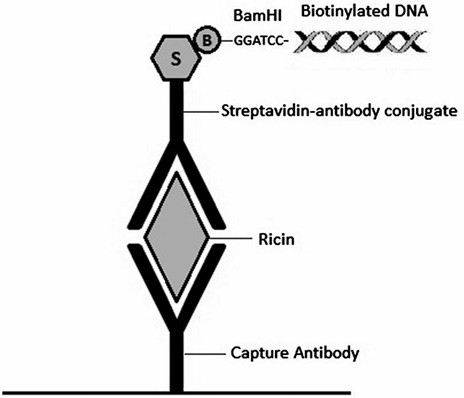
Figure 1. Schematic representation of the Sandwich IPCR method, depicting the analytical complex on the surface of an assay well.

IPCR offers several advantages over conventional protein detection methods. First, the limit of detection is greatly improved compared to immunoassays. Second, the sample volume required is very small. The high sensitivity of IPCR enables the analysis of sample sizes of less than 1 μL. This is of particular importance for studies where only limited volumes of samples are available. Third, the assay is compatible with most complex biological matrices. Owing to the high sensitivity of IPCR, the biological sample can usually be diluted, which significantly reduces the matrix effect. Fourth, the use of real-time PCR, rather than end point PCR, improves the quantitative accuracy of the assay.

Here, we introduce a sandwich IPCR assay. This assay uses a "sandwich" of antibodies to capture and detect a protein of interest as done in a sandwich ELISA, with an additional step using a DNA marker that binds to the detection antibody through an avidin–biotin interaction allowing for signal amplification by realtime PCR.

View all ELISA Positive Control

View all ELISA Matched Antibody Pair

The method takes advantage of the high affinity bond between biotin and streptavidin to form a streptavidinated antibody-biotinylated DNA complex. Because excess proteins and DNA can interfere with PCR, the DNA is made cleavable from the antibody complex by incorporating a BamHI restriction site at the 5' end. After cleaving the DNA from the immuno-complex, an aliquot is transferred to real-time PCR tubes or 96-well plates containing a PCR master mix cocktail with primers and a probe with a fluorescent reporter. Throughout the PCR cycle, the real-time PCR cycler detects and records changes in the fluorescence signal during amplification. This amplification signal is used to calculate the cycle threshold (Ct) value. The Ct is defined as the number of cycles required for the fluorescent signal to cross the threshold (i.e., exceed background levels). Ct levels are inversely proportional to the amount of DNA in the sample such that the lower the Ct value, the greater the amount of DNA in the sample and, therefore, the greater the amount of target antigen. The DNA probe has a fluorescent reporter at one end and a quencher of fluorescence at the opposite end of the probe. The close proximity of the reporter to the quencher prevents detection of its fluorescence until the 5' to 3' exonuclease activity of the Taq polymerase breaks the reporter–quencher proximity and thus allows emission of fluorescence, which can be detected upon excitation with a laser. An increase in the product targeted by the reporter probe at each PCR cycle therefore causes a proportional increase in fluorescence due to the breakdown of the probe and release of the reporter.

As with all immunoassays, the sensitivity of IPCR largely depends on the antibodies selected to bind and detect the target antigen. The sandwich IPCR requires two antibodies, each with a strong affinity to different epitopes of the antigen. A polyclonal antibody can be used as both the capture and detection antibody but the use of a monoclonal antibody for capture tends to provide more reproducible results due to the consistent and even coating across each well. It is recommended that sandwich ELISAs be carried out to determine the best antibody pair for each particular antigen before applying the antibodies to the IPCR method. The limit of detection (LOD) for IPCR is defined as the average Ct value of the negative controls (wells containing buffer or sample matrix only without any antigen) plus three times the calculated standard deviation. In order to calculate the standard deviation and, subsequently, the LOD, it is necessary to perform the assay with triplicates for each sample.

Using the procedures described here, we demonstrate that the sandwich IPCR can detect 10 pg/mL of ricin in chicken egg and bovine milk samples and 100 pg/mL in ground beef extracts. Comparable ELISA results were in the 1–10 ng/mL range. Thus, IPCR affords sensitivity that is tenfold greater in the ground beef matrix, 100-fold greater in the milk matrix, and 1,000-fold greater in the egg matrix than the sensitivity obtained by ELISA. This IPCR is also highly compatible with complex environmental matrices. When applied to 23 environmental samples including feces, feral swine colon, soil, and water from watersheds for detecting the presence of Shiga toxin 2 produced by Shiga toxin-producing E. coli (STEC), it demonstrated a 100 % sensitivity and specificity.

Materials of Immuno-PCR Assay Protocol

Prepare all solutions using ultrapure water (prepared by purifying deionized water to attain a sensitivity of 18 MΩ cm at 25 °C) and analytical grade reagents. Prepare and store all reagents at room temperature (unless otherwise indicated). Follow all waste disposal regulations when disposing of waste materials.

  1. Reagent buffer: 50 mM boric acid, pH 9.5. Weigh out 0.309 g boric acid and add water and 1–5 M NaOH to 100 mL volume at pH 9.5. Sterilize by filtration and store in 5–10 mL aliquots at − 20 °C.
  2. Tris-buffered saline (TBS): 20 mM Tris-Cl, 150 mM NaCl, pH 7.5. Weigh out 8 g NaCl, 0.2 g KCl, and 3 g Tris Base and dissolve into 1 L water plus 1–5 M HCl to pH 7.5. Autoclave at 121 °C for at least 30 min to sterilize.
  3. Bovine Serum Albumin–Tris-buffered saline (BSA–TBS): TBS containing 0.5 % (wt/vol) bovine serum albumin (BSA), 5 mM EDTA, and 0.2 % (wt/vol) NaN3. Dissolve 1 g BSA, 0.4 g NaN3, and 0.29 g EDTA into 200 mL TBS (see Note 1). Sterilize by filtration and store in 50 mL aliquots at −20 °C (see Note 2).
  4. Tween/EDTA/Tris-buffered saline (TETBS): TBS containing 5 mM EDTA and 0.05 % (vol/vol) Tween 20. Weigh out 1.46 g EDTA and stir into 1 L TBS until dissolved completely. Stir in 0.5 mL Tween 20 until dissolved completely (see Note 3). Autoclave at 121 °C for at least 30 min to sterilize.
  5. Reagent dilution buffer: Prepare from fresh BSA–TBS and TETBS in a 1:10 ratio. Mix 1 mL BSA–TBS with 9 mL TETBS (see Note 4).
  6. Anti-ricin monoclonal antibody 1642.
  7. Streptavidin-conjugated anti-ricin antibody: prepare using Lightning-Link Streptavidin Conjugation Kit following the manufacturer's instructions: Mix 100 μL of 1 mg/mL anti-ricin polyclonal goat antibody (Vector Laboratories, Burlingame, CA) with 10 μL of LL-Modifier reagent and then add to vial containing 100 μg of lyophilized LL-streptavidin. Incubate the mixture for 3 h at room temperature (RT), and then add 10 μL of LL- quencher reagent. The conjugate can be used after 30 min or stored at 4 °C.
  8. Biotinylated DNA marker: A mono-biotinylated DNA marker was prepared by PCR using the pUC19 plasmid as the template and the primer pair: pUC-bio (20) (5'-biotin-CCCGGATCCCAGCAATAAACCAGCCAGCC- 3') and F1 (5'-TAT GCAGTGCTGCCATAACCATGA- 3'). Perform amplification with Taqman Universal PCR Mastermix with an initial 95 °C denaturation for 10 min followed by 40 cycles of 95 °C for 15 s and 60 °C for 1 min (see Note 5).
  9. BamHI restriction endonuclease and buffer 3.1.
  10. 96-well V-bottom microtiter plate (see Note 6).
  11. Adjustable speed orbital plate shaker such as the Barnstead/ Lab-Line Titer Plate Shaker, or similar.
  12. Incubator set to 37 °C.
  13. Multichannel pipette and filter tips (see Note 7).
  14. 1.5 mL low-adhesion microfuge tubes (see Note 8).
  15. 0.2 mL Temp Assure PCR 8-tube flex-free strips with individually attached, optically clear flat caps or similar Real-time PCR tubes or plates (see Note 9 ).
  16. TaqMan Universal PCR 2× Mastermix
  17. Primers: F (5'-CCATAACCATGAGTGATAACACTGCT-3') R (5'-CGATCAAGGCGAGTTACATGATC-3')
  18. Probe (5'-Fam-ACCGAAGGAGCTAACCGCTTTTTTGC AC- Tam-3')
  19. Real-Time PCR cycler (see Note 10). 

Methods of Immuno-PCR Assay Protocol

Carry out all procedures at room temperature unless otherwise specified. Plate washes are carried out on an oscillating plate shaker, set to a low shaking speed (see Note 11). Wear gloves for all steps and change them frequently to avoid contamination. Keep workspace clean and free of DNA, antigens, and other sources of contamination by wiping down regularly.

Immunoassay

  1. Dilute anti-ricin monoclonal antibody 1642 to 4 μg/mL in reagent buffer and coat a 96-well microtiter plate with 30 μL of antibody dilution per well (see Note 12).
  2. Incubate covered plate overnight at 4 °C (see Note 13).
  3. Wash the wells three times for 1 min with 150 μL of TBS (see Note 14).
  4. Block non-adsorbed sites with 150 μL of BSA–TBS per well and incubate for 1 h (see Note 15).
  5. Wash wells twice for 30 s and twice for 4 min with 150 μL of TETBS.
  6. Prepare tenfold serial dilutions of ricin in TBS and add 30 μL per well of each dilution and incubate for 1 h (see Note 16).
  7. Wash wells as in step 5.
  8. Add 30 μL per well of streptavidin-conjugated anti-ricin polyclonal antibody at a final concentration of 80 ng/mL in reagent dilution buffer and incubate at 37 °C for 30 min.
  9. Wash wells as in step 5.
  10. Add 30 μL per well of biotinylated DNA marker at 0.5 ng/μL in TETBS and incubate for 30 min. Keep any remaining dilution on ice for the positive PCR control.
  11. To remove unbound DNA, wash wells four times for 30 s and three times for 4 min with TETBS, followed by fi ve times for 1 min with TBS.
  12. Detach bound DNA from the immuno-complex by incubating the wells with 30 μL of restriction buffer containing 1 unit per well of BamHI for 2 h at 37 °C ( see Note 17 ).
  13. Use 6 μL of digested DNA as a template in real-time PCR (20 μL reaction volume) (see Note 18).

Real-Time PCR

  1. Prepare 1× PCR mastermix as follows, multiplying the volume by the number of samples to be analyzed (see Note 19):
    1. TaqMan Master Mix (2×) 10 μL
    2. 10 μM primer F 0.6 μL
    3. 10 μM primer R 0.6 μL
    4. 10 μM Probe 0.5 μL
    5. Water 2.3 μL
  2. Aliquot 14 μL PCR mastermix to real-time PCR tubes with caps, one per sample, plus one each for positive and negative PCR controls.
  3. Add 6 μL of digested DNA as a template. For the positive control sample, add 1 μL of the DNA marker dilution at 0.5 ng/μL and 5 μL of water. For the negative control, add 6 μL of water to the master mix. Close all caps. Wipe caps with precision wipes to remove dust before placing in PCR cycler (see Note 20).
  4. Run PCR program: 95 °C 10 min (95 °C 15 s, 62 °C 1 min), ×40 cycles, 4 °C, hold. Use a heated lid. Set the volume to 20 μL and background calibration to IPCR tubes (see Note 21). Set FAM as the fluorophore and tetramethylrhodamine as the quencher.
  5. Use Realplex software to calculate baseline and cycle threshold (Ct) values (see Note 22).

Notes of Immuno-PCR Assay Protocol

  1. BSA–TBS includes the preservative NaN3 which can cause harm via contact with skin, eyes or upon ingestion. Use proper PPE when preparing and handling this solution.
  2. BSA–TBS can be frozen and thawed repeatedly but should remain on ice throughout the assay procedure.
  3. Tween is very viscous and sticky and therefore needs to be measured precisely and dissolved completely into solution to get an accurate final concentration.
  4. Reagent dilution buffer should be made fresh and kept on ice during the assay and any excess discarded after use.
  5. A BamHI restriction site was included in the pUC-bio primer to allow removal of the DNA marker from the immunocomplex. The resulting PCR product should be about 340 bp when run on an agarose gel for analysis. Purify the PCR product using a QIAquick PCR Purification Kit and determine the concentration by Nanodrop or using a UV–visible spectrophotometer.
  6. You may substitute TopYield strips or other thermally stable, small-welled plate with a high-binding capacity. If using Top Yield strips, increase the wash and block buffer volumes to 200 μL per well.
  7. Filter tips should be used to prevent cross-contamination. Be careful not to touch the tips to the wells of the plate while pipetting and change them between each washing and between pipetting different reagents or antigens into the wells.
  8. For preparation of antigen dilutions, low adhesion tubes are used to minimize nonspecific binding of protein, allowing greater accuracy and sensitivity when performing IPCR.
  9. If using a PCR plate, it must be sealed with optically clear tape and covered with a compression pad (rubber mat with holes, aligned properly for measurement of fluorescence) when placed in the PCR cycler.
  10. PCR can also be done using a conventional PCR cycler followed by analysis of DNA concentrations by gel electrophoresis. However, quantitative real-time PCR has the advantages of shorter handling time and greater reproducibility.
  11. The shaker should be set to a speed of two or three, such that the plate is oscillating swiftly but the wash solution is not splashing from the wells. Alternatively, an automatic plate washer may be used with the following protocol: aspirate, wash 6 × 150 μL, with aspiration following each wash.
  12. The concentration of capture antibody for coating plates can vary but should be in the range of 0.5–5 μg/mL for best results.
  13. Cover with plastic wrap or plate-sealing tape to prevent evaporation from wells. Plates can be left at 4 °C for up to 3 days with little or no effect on assay outcome.
  14. For each wash step, use a multichannel pipette to add wash buffer to wells, then place on the orbital shaker for the time specified. Repeat as specified.
  15. Alternate blocking buffers/concentrations can be substituted if high background is observed in a particular application of IPCR that may not be compatible with BSA or requires a higher concentration of blocking agent.
  16. The dilutions should be in the range of 0.01 pg/mL to 100 ng/mL. The optimal range will vary based on the strength of antibodies used. When analyzing samples with unknown antigen concentration, be sure to include a full set of standards for comparison and quantification.
  17. Use a buffer suitable for BamHI, such as buffer 3.1 or 2.1 from New England BioLabs, and dilute it to 1× with sterile nuclease-free water prior to adding the restriction endonuclease. The endonuclease typically is supplied at a concentration of 20 units/μL and should be diluted to yield 1 unit per 30 μL. Cover plate with a tight-sealing tape or film to prevent evaporation.
  18. Remaining digested DNA can be kept at 4 °C overnight if the experiment needs to be repeated the next day.
  19. For short-term storage (less than 30 min), keep mastermix on ice in the dark to protect the probe against degradation.
  20. Any dust particles or other contamination on the PCR tubes will block the measurement of fluorescence and skew the results.
  21. Follow instructions in the real time PCR cycler manual for calibrating the tubes or plates that will be used for the PCR step.
  22. If random signals and high error are observed, check PCR wells for background fluorescence and contamination. Also check PCR tubes for evaporation and ensure heated lid is working. If positive signals are observed in the negative controls, check all PCR reagents for contamination.

Reference

  1. Robert Hnasko. ELISA Methods and Protocols. Methods in Molecular Biology. 2015, 1318. Electronic ISSN: 978-1-4939-2742-5.
Inquiry Basket