
Inquiry Basket
 Loading ......
Nipah virus (NiV) is a highly pathogenic paramyxovirus that emerged in 1998 from fruit bats in Malaysia. It caused severe respiratory disease in pigs and fatal encephalitis in humans with high mortality rates. Unlike other paramyxoviruses, NiV can infect a wide range of mammalian species. Due to its broad host range, zoonotic potential, high pathogenicity, and lack of effective vaccines or treatments, NiV is classified as a biosafety level 4 pathogen. The glycoproteins of the Nipah virus, especially two surface glycoproteins: the receptor-binding G protein and the fusion protein F, are major determinants for cell tropism and virus spread. Understanding these glycoproteins is crucial for understanding viral pathogenicity.
The presence of two specific surface glycoproteins, G and F, in the NiV is crucial for the virus to enter host cells, determining its ability to infect a broad range of species and different types of cells. The G protein helps the virus attach to specific receptors on the surface of host cells, while the F protein initiates the fusion of the viral membrane with the cellular membrane, allowing the virus to enter the host cell. When both G and F proteins are present, either on infected cells or cells expressing the NiV glycoproteins, the F protein facilitates fusion between adjacent cells, leading to the formation of multinucleated syncytia, which are clusters of fused cells. This process of cell-to-cell fusion allows the virus to spread directly from infected cells to uninfected neighboring cells without the need for virus budding. The presence of syncytia formation significantly impacts the damage caused by NiV infection and the progression of the disease.
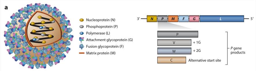 Figure 1. Structure and genome organization of NiV.
(Source: Amaya, M. et al., 2020)
The G protein of the NiV is a type II membrane glycoprotein that consists of 602 amino acids. Its primary function is to bind to receptors on host cells. In addition to receptor binding, the G protein is crucial for facilitating the fusion of lipid membranes through the action of the F protein. Unlike most other paramyxoviruses, the NiV G protein does not possess hemagglutinating or neuraminidase activities, and it does not bind to carbohydrate structures.
Recent research has confirmed that NiV utilizes a cellular surface protein called ephrin B2 as its receptor. Ephrin B2 is a glycoprotein normally involved in cell signaling during development, particularly in the vascular and nervous systems. It is widely expressed in various cells and tissues, with high levels found in smooth muscle cells, neurons, and endothelial cells. Ephrin B2 is also upregulated in certain tumor cells, explaining the broad cell tropism of NiV in cultured tumor cells. Additionally, ephrin B3 has been suggested as an alternative receptor for NiV, potentially contributing to the neurological symptoms observed in NiV patients in regions where ephrin B2 is absent.
The F protein of Nipah virus (NiV) is a type I transmembrane protein consisting of 546 amino acids. It undergoes glycosylation and oligomerization into trimers in the endoplasmic reticulum. The F protein is then transported to the plasma membrane via the Golgi apparatus. Before being incorporated into new virions, the precursor F0 protein is cleaved into two subunits, F1 and F2. The F1 subunit contains functional domains, including a cytoplasmic tail, a transmembrane domain, and a fusion peptide at the N terminus. The fusion peptide plays a critical role in initiating the fusion process by inserting into the target membrane. The F1 subunit also contains heptad repeats, which form a 6-helix bundle structure during membrane fusion. Peptides corresponding to the C-terminal heptad repeat of the NiV F protein have been shown to inhibit NiV-mediated membrane fusion.
N-glycosylation plays a significant role in the structural and functional aspects of fusion proteins. It affects their folding, transport, activity, and immunological properties. The NiV F protein, along with the closely related Hendra virus (HeV) F protein, contains potential N-glycosylation sites. Analysis reveals that specific sites in both F1 and F2 subunits are utilized for N-glycosylation. These glycans vary in complexity and type. The glycan at N414 is crucial for protein folding and cell surface transport, while the absence of the oligosaccharide attached to N464, which lies within the heptad repeat-2, does not significantly affect fusion activity. This differs from other paramyxoviral fusion proteins where the glycan within the heptad repeat is essential for fusion. Recent studies have shown that N-glycosylation of the F protein also influences its association with the G protein and affects the fusogenicity of the NiV glycoprotein complex. Furthermore, N-glycans are thought to protect NiV from antibody neutralization, but their precise role in modulating antigenicity is yet to be fully understood.
Proteolytic cleavage of the fusion protein in paramyxoviruses is necessary for virus infectivity. The distribution of proteases that activate the fusion protein plays a crucial role in determining the host range, tissue tropism, and pathogenicity of the virus. There are two known mechanisms of fusion protein activation: intracellular cleavage by the Golgi protease furin or extracellular cleavage by trypsin-like proteases. NiV has a single basic amino acid residue at the cleavage site, indicating that its activation mechanism is different from other paramyxoviruses. NiV replication depends on a cellular protease and occurs in the absence of exogenous trypsin. Activation of NiV F proteins takes place at the cell surface through endocytosis and proteolytic processing in the acidic environment of endosomes. The cytoplasmic tail of the NiV F protein contains an endocytosis signal that ensures constitutive internalization and cleavage. Moreover, the involvement of the endosomal protease cathepsin L in cleaving NiV F proteins further highlights their distinct activation mechanism compared to other paramyxoviruses.
Reference
| Target | Cat. No. | Product Name | Expression System | Tag/Conjugate | Application | |
| Nipah Virus | DAG-WT1059 | Recombinant Nipah Virus Glycoprotein G | Insect cells | Unconjugated | Immunoassays | Inquiry |
| DAG-WT633 | Recombinant Nipah Virus F Protein [His] | HEK293 | His | ELISA | Inquiry | |
| DAG-WT634 | Recombinant Nipah Virus G Protein [His] | HEK293 | His | ELISA | Inquiry | |
| DAG-WT271 | Recombinant Nipah Virus G Protein [Fc] | HEK 293 | Fc | SDS-PAGE | Inquiry | |
| DAG-WT272 | Recombinant Nipah Virus F Protein [Fc] | HEK 293 | Fc | SDS-PAGE | Inquiry | |
| DAG-WT273 | Recombinant Nipah Virus Nucleoprotein [His] | HEK 293 | His | ELISA | Inquiry | |
| DAGA-1004 | Recombinant Nipah Virus G Protein | Insect cells | TBD | ELISA | Inquiry | |
| DAG-WT381 | Recombinant Nipah Virus (NiV) VLP | HEK293 | Unconjugated | Immunoassays | Inquiry |
| Target | Cat. No. | Product Name | Size | Species Reactivity | Application | Detection Sample | |
| FURIN | DEIA10900 | Human FUR(Furin) ELISA Kit | 96T | Human | Quantitative | Serum, plasma, tissue homogenates and other biological fluids. | Inquiry |
| DEIA5404 | Human Furin ELISA kit | 96T | Human | Quantitative | serum, plasma, cell culture supernatant, urine | Inquiry |
Loading ......
