Nipah virus F protein, recombinant protein from HEK293 cells
Enzyme-Linked Immunosorbent Assay Using Henipavirus-Receptor EphrinB2 and Monoclonal Antibodies for Detecting Nipah and Hendra Viruses
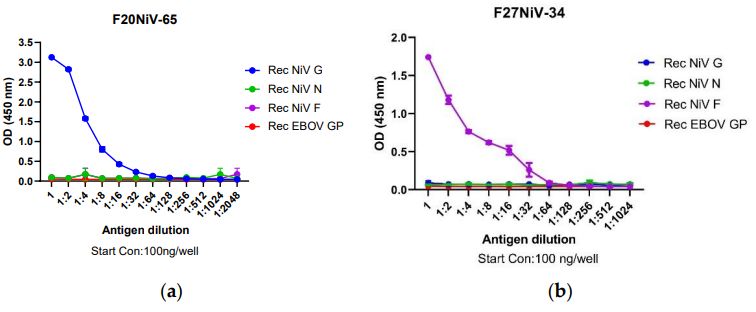 Figure 1. Reactivity of mAbs to recombinant NiV structural proteins in indirect ELISA.
 Development of a Point-of-Care Immunochromatographic Lateral Flow Strip Assay for the Detection of Nipah and Hendra Viruses
 Figure 1. Reactivity of monoclonal antibodies against inactivated NiV/HeV and recombinant NiV structural proteins
 | Product Name | Cat. No. | Applications | Host Species | Datasheet | Price | Add to Basket |
|---|
| Product Name | Cat. No. | Applications | Host Species | Datasheet | Price | Add to Basket |
|---|
Commonly found in Southeast Asia, Nipah virus (NiV) was first discovered in Malaysia in 1998 and is considered one of the world's deadliest viruses with a very high mortality rate (40%-70%). NiV is classified as a biosafety class 4 (BSL-4) pathogen. It is a zoonotic RNA virus, belonging to the genus Henipavirus from the Paramyxoviridae classification, with a size of 40nm-1900nm. Compared with typical paramyxovirus, the composition of NiV is slightly different. NiV contains reticular cytoplasmic inclusion, and its average volume is larger than that of most paramyxoviruses. Many paramyxoviruses have the characteristics of hemagglutinin and neuraminidase, while NiV does not. NiV enters the target cell through microphagocytosis of G protein and F protein, and its transcription and replication pathways are similar to that of other paramyxovirus. Primary transcription includes the RNA polymerase complex packaged in the virus, which replicates the viral RNA and then produces capped, short capped, uncapped RNA and polyadenylated mRNA that encode the viral protein.
 Figure 1. Schematic representation of the structure of Nipah Virus (NiV) and the organization of the genome
(Source: Alaskari M, et al. 2023)
NiV causes mainly acute encephalitis and respiratory disease with high mortality. A small number of infected individuals have no symptoms. The incubation period is 4-21 days, followed by prodromal symptoms and clinical manifestations of fever, headache and myalgia. The symptoms of encephalitis appear within a week and are usually characterized by a change in mental status, blunted reflexes, low intraocular pressure, segmental myoclonus, paralysis of the gaze, and weakness of the limbs. The patient's condition deteriorates rapidly, with coma and death occurring rapidly within a few days. 20% of survivors have residual neurological deficits, including fatigue, focal neurological deficits, and depression.
NiV can be diagnosed by molecular and serological detection, virus isolation, histopathology, and immunohistochemistry. The main detection methods are real-time reverse transcription polymerase chain reaction (rRT-PCR) for nucleic acid and enzyme-linked immunosorbent assay (ELISA) for antibody detection. rRT-PCR detection is faster, more specific, and more sensitive, so it is the first choice for the diagnosis of NiV infection. ELISA is the most commonly used serological diagnostic test with high sensitivity, simple and safe use, and can monitor the condition of patients and animals.
NiV F Protein
References
1. Alaskari M, et al. Nipah Virus: A Threatening Outbreak. Journal of Clinical and Diagnostic Research. 2023 Feb; 17. DE01 - DE07.
2. Aditi, et al. Nipah virus infection: A review. Epidemiol Infect. 2019 Jan;147:e95.
Q: What strain of Nipah is the F sequence from? Is it Malaysia or Bangladesh?
A: NiV-M (Malaysia)
Q: We recently purchased a recombinant protein, DAG-WT633. The product sheet did not include a recommended buffer for resuspension of the lyophilized product. Our intended downstream application is ELISA, for which we typically use PBS as the coating buffer. Would it be acceptable to resuspend the protein in PBS? Although the protein will be further diluted in this buffer, we wanted to confirm before proceeding.
A: • Reconstitution
It is strongly recommended to reconstitute the lyophilized Nipah virus Pre-Fusion glycoprotein, His Tag with 1 250 µL sterile deionized water to obtain a 400 µg/mL stock solution. Allow the protein to solubilize for 30–60 minutes at room temperature with occasional gentle mixing. Avoid vigorous shaking or vortexing.
To prevent surface-adsorption loss and inactivation, the reconstituted protein should not be aliquoted to less than 10 µg per vial.
• ELISA dilution
Once reconstituted, the protein can be further diluted in PBS for ELISA coating without any issues.
Please let us know if you need additional information.
ChAdOx1 NiV vaccination protects against lethal Nipah Bangladesh virus infection in African green monkeys
NPJ Vaccines
Authors: van Doremalen N, Avanzato VA, Goldin K, Feldmann F, Schulz JE, Haddock E, Okumura A, Lovaglio J, Hanley PW, Cordova K, Saturday G, de Wit E, Lambe T, Gilbert SC, Munster VJ.
Immunological correlates of protection afforded by PHV02 live, attenuated recombinant vesicular stomatitis virus vector vaccine against Nipah virus disease.
Front Immunol
Authors: Monath TP, Nichols R, Feldmann F, Griffin A, Haddock E, Callison J, Meade-White K, Okumura A, Lovaglio J, Hanley PW, Clancy CS, Shaia C, Rida W, Fusco J.
![]()
