
Inquiry Basket
 Loading ......
Lysosomes were first discovered in rat liver cells. Subsequent studies found that various biological macromolecules in cells are mainly degraded in lysosomes. A distinctive feature of lysosomes is their acidic internal environment (pH value between 4.5 and 5.5). The acidic environment is conducive to maintaining the activity of a large number of hydrolases (including proteases, glycosidases, lipases, etc.) in lysosomes. Cells use a variety of pathways, including phagocytosis, macropinocytosis, and autophagy, to deliver extracellular biological macromolecules, intracellular protein aggregates, and damaged organelles into lysosomes, undergo degradation.
The powerful degradation effect of lysosomes is very important for the cleanup of intracellular toxic substances, removal of damaged organelles, regulation of signal transduction, and maintenance of the intracellular environment. If the genes encoding lysosomal hydrolases or permeases are mutated, affecting the normal functions of these proteins, a type of genetic disease called lysosomal storage disease (LSD) often occurs. The functional decline of liver and other organs caused by aging is also closely related to the decline of lysosomal function. Rapidly dividing tumor cells need to use the degradation function of lysosomes to cope with changes in the tumor microenvironment and lack of nutrients.
A large number of studies in recent years have fundamentally changed the previous inherent views on lysosomes, showing that lysosomes are not only the degradation and recycling station for various metabolic wastes in cells, but also an important regulatory point for complex intracellular signaling networks.
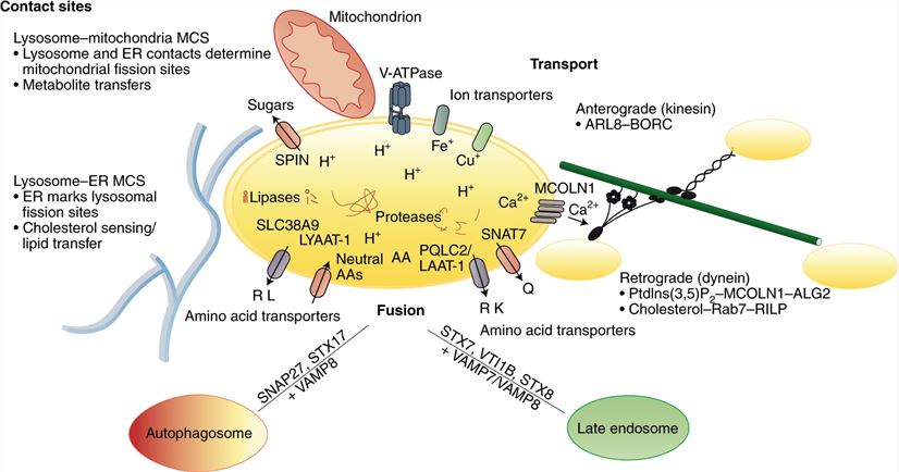 Figure 1. Lysosome cell biology. (Lawrence RE, et al.; 2019)
Lysosomes are present in all eukaryotic cells, but the lysosomes contained in different cells vary greatly in shape, size, number, and function. Most mammalian cells contain hundreds of lysosomes with a diameter of 0.1 to 1 μm, while yeast and plant cells contain one or several vacuoles similar to lysosomes. Lysosomes are organelles wrapped in a single membrane, and a large number of transmembrane proteins are embedded in the lysosomal membrane. Most lysosomal membrane proteins located in the lysosomal lumen are highly glycosylated to protect them from degradation by proteolytic enzymes within the lysosome. In recent years, lysosomal proteomics analysis has discovered some new lysosomal proteins with unknown functions, which may be involved in the degradation of biological macromolecules, the transport of metabolites, and signal transduction.
Lysosomes mainly originate from the intracellular vesicle transport system. Clathrin-mediated endocytosis at the cell membrane wraps extracellular cargo into vesicles to form early endosomes. Early endosomes fuse with vesicles derived from the Golgi apparatus to generate late endosomes/multivesicular bodies (MVBs). Multivesicular bodies are characterized by a vesicle structure formed by the invagination of multiple vesicles inside, which was first discovered to play a role in capturing and degrading epidermal growth factor (EGF). Follow-up studies found that multivesicular bodies can also accept cargo from the trans-Golgi network (TGN) or re-deliver cargo to the trans-Golgi network. In addition to being transformed into late endosomes/multivesicular bodies, early endosomes can also be transported back to the cell membrane or divide to produce recycling endosomes. Recycling endosomes can also be transported back to the cell membrane or retrogradely transported to the reverse Golgi apparatus. The generation process of lysosomes is an integral part of the intracellular vesicle transport system. The transformation process from early endosomes to late endosomes is accompanied by the gradual acidification of the luminal environment, originating from the Golgi apparatus carrying a complete set of lysosomal hydrolysis Vesicles of enzymes and other lysosomal proteins fuse with acidified late endosomes, eventually forming mature lysosomes with powerful degradation capabilities. Research in recent years has found that intracellular lysosomes can also be produced through autophagic lysosome reformation (ALR). In the late stage of the autophagy pathway, autophagic lysosomes fuse with lysosomes to form autophagic lysosomes. The body can form lysosomes by separating out lysosome-specific components.
The movement of lysosomes within cells mainly occurs on microtubules. Lysosomes can rely on kinesin to move from the positive pole of microtubules to the negative pole of microtubules, and they can also rely on dynein to move from the negative pole of microtubules to the positive pole of microtubules. Lysosomes can frequently and rapidly change their movement direction within cells, but it is still not clear what factors regulate the movement rate and direction of lysosomes. Current research shows that it may be related to the acidification of lysosomes and cell migration. Closely related to the balance of nutrients within cells.
In recent years, studies have found that a large number of genes encoding lysosomal proteins (including hydrolases, permeases, membrane proteins, etc.) can be regulated by some specific transcription factors at the same time. Bioinformatics analysis found that there are base sequences called coordinated lysosomal expression and regulation (CLEAR) elements in the promoter regions of many lysosomal genes. Genes with CLEAR elements in their promoter regions are generally regulated by a family of transcription factors called MiT/TFE (microphthalmia/transcription factor E) proteins. The main members of the MiT/TFE protein family include transcription factor EB (TFEB), transcription factor EC (TFEC), transcription factor E3 (TFE3) and microphthalmia-associated transcription factor (MITF). TFEB and other members of the MiT/TFE protein family can directly bind to the CLEAR element and promote the expression of downstream target genes. Interestingly, TFEB also promotes the expression of a large number of autophagy-related proteins. TFEB can significantly enhance the degradation efficiency of lysosomes by coordinating the promotion of autophagy and lysosomal biogenesis.
Autophagy is a general term for a self-digestion pathway unique to eukaryotes that degrades intracellular protein aggregates and damaged organelles. There are three main forms of autophagy pathways in eukaryotic cells: macroautophagy, chaperone-mediated autophagy, and microautophagy. The process by which molecular chaperone proteins recognize and bind to specific amino acid sequences on substrate proteins and transport the substrate proteins to lysosomes for degradation is called chaperone-mediated autophagy. The process in which mammalian late endosomes/lysosomes or yeast vacuoles use membrane invaginations to directly wrap substrates for degradation is called microautophagy. When macroautophagy occurs, vesicles with a double-membrane structure called autophagosomes will first be formed in the cell. During the formation process, the autophagosomes will wrap up the substances to be degraded. Enzyme fusion transports substrates to lysosomes for degradation. Therefore, the above three forms of autophagy pathways ultimately strictly rely on lysosomes for degradation.
When the cytoplasmic membrane is damaged, high concentrations of extracellular calcium ions quickly enter the cytoplasm through the broken pores in the cytoplasmic membrane, causing the intracellular calcium ion concentration to increase. The repair process of the cytoplasmic membrane requires the participation of calcium ions, and the exocytosis process induced by calcium ions has been considered to possibly play a role in the repair process of the cytoplasmic membrane. Research in recent years has found that calcium ions entering the cytoplasm from the damaged cytoplasmic membrane will trigger the exocytosis of lysosomes. The vesicles carrying the lysosomal membrane will block the damaged pores on the surface of the cytoplasmic membrane by fusing with the damaged cytoplasmic membrane. Thereby restoring the integrity of the cell plasma membrane.
References
| Target | Cat. No. | Product Name | Expression System | Tag/Conjugate | Application | |
| CTNS | DAG-WT364 | Recombinant Human CTNS Protein [GST] | Wheat Germ | GST | WB/ELISA/SDS-Page | Inquiry |
| LAMP1 | DAG-P2064 | LAMP1 peptide | N/A | Unconjugated | ELISA | Inquiry |
| DAG-P1739 | Human LAMP1 blocking peptide | N/A | Unconjugated | BL | Inquiry | |
| DAG-P1740 | Human LAMP1 blocking peptide | N/A | Unconjugated | BL | Inquiry | |
| CDBP5661 | LAMP1 blocking peptide | N/A | Unconjugated | IB | Inquiry | |
| LAMP2 | DAG-P0773 | Human LAMP2 peptide | N/A | Unconjugated | ELISA | Inquiry |
| DAG-P0774 | Human LAMP2 blocking peptide | N/A | Unconjugated | BL | Inquiry | |
| DAG-P0775 | Human LAMP2 blocking peptide | N/A | Unconjugated | BL | Inquiry | |
| CDBP5662 | Lamp2 blocking peptide | N/A | Unconjugated | IB | Inquiry | |
| DAG-KO200 | LAMP2 Knockout Cell Lysate | WB | Inquiry |
| Target | Cat. No. | Product Name | Size | Species Reactivity | Application | Detection Sample | |
| LAMP1 | DEIA-FN785 | Mouse LAMP1 (Lysosomal Associated Membrane Protein 1) ELISA Kit | 96T | Quantitative | serum, plasma, cell culture supernatants, tissue homogenate | Inquiry | |
| DEIA-FN786 | Human LAMP1 (lysosomal-associated membrane protein 1) ELISA Kit | 96T | Quantitative | serum, plasma, cell culture supernatants, tissue homogenate | Inquiry | ||
| ABPR-ZB195 | Human LAMP1 Antibody Pair Set | 5 Plates, 15 Plates | Human | sELISA | Inquiry | ||
| LAMP2 | DEIA-FN787 | Human LAMP2 (Lysosome-associated membrane glycoprotein 2) ELISA Kit | 96T | Quantitative | serum, plasma, cell culture supernatants, tissue homogenate | Inquiry | |
| DEIA-LL053 | Mouse LAMP2 ELISA Kit | 96T | Mouse | Quantitative | Serum, plasma, tissue homogenates and other biological fluids | Inquiry | |
| LAMP3 | ABPR-ZB291 | Human LAMP3/CD208 Antibody Pair Set | 5 Plates, 15 Plates | Human | sELISA | Inquiry |
Loading ......
