
Inquiry Basket
 Loading ......
Androgens are a steroid hormone that defines male sexuality and controls secondary male characteristics. They're involved in male sex development and spermatogenetic growth and regulation. They also make the male organs, and start and maintain spermatogenesis. Androgens — testosterone and dihydrotestosterone (DHT) — both go to the androgen receptor, but they aren't identical. Testosterone directs development of largely structures born from the Wolffian duct: the epididymis and seminal vesicles; DHT governs the formation of outer genitalia like the prostate gland, scrotum and penis. The body releases testosterone in 3 forms and 5-alpha-reductase, which is a mechanism used in target cells to convert testosterone into DHT, binds through activation of the androgen receptor (AR).
Androgens start their production with cholesterol, converted into testosterone in the testes' Leydig cells. This is initiated as early as the sixth week of gestation, and initially doesn't depend on LH. But, as testis grows, LH from the pituitary gland skews testosterone levels. LH is modulated by the hypothalamus through gonadotropin-releasing hormone (GnRH), which testosterone negative feedbacks to inhibit.
Conversion of cholesterol to testosterone is multi-step process. It first proceeds through steroidogenic acute regulatory protein (StAR), which makes cholesterol travel to mitochondria. HP450scc here side-chains cholesterol into pregnenolone, the lowest energy androgen biosynthesis step. Then it goes on to enzymes like 3beta-hydroxysteroid dehydrogenase and 17beta-hydroxysteroid dehydrogenase type 3. Or testosterone, converted into DHT by 5-reductase type 2, a permanent testosterone-to-DHT conversion enzyme. It is the conversion responsible for male external genitalia such as the penis and scrotum to be differentiated. Recent research has also highlighted an alternative "backdoor" pathway for DHT production, which plays a role in fetal masculinization and certain disorders like congenital adrenal hyperplasia.
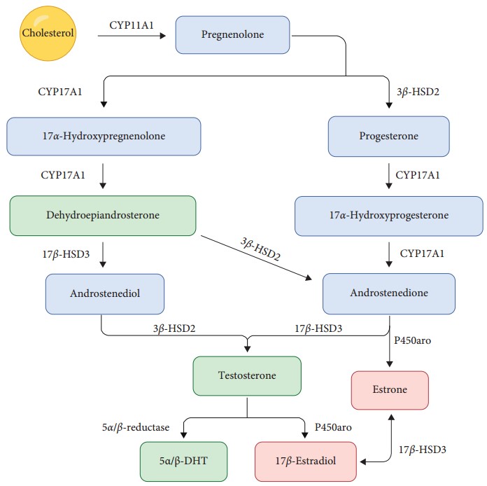 Figure 1. Androgen synthesis from cholesterol
(Source: Carbajal-García A, et al. 2020)
Androgens biological effects operate through their interaction with AR. Testosterone (or DHT) attaches to AR in the cells targeted, which triggers molecular cascades that change gene expression. Armed with its ligand, AR changes structure and dissociates from chaperone proteins (i.e., heat shock proteins), which contain AR as a ghost. The activated AR then translocates to the nucleus where it binds to specific DNA sequences known as androgen response elements (AREs).
Once bound to DNA, AR forms a homodimer and recruits additional proteins, including coactivators and general transcription factors. This complex facilitates transcriptional activation or repression of androgen-responsive genes, leading to cellular changes that govern male sexual differentiation and secondary characteristics.
Interestingly, androgen signaling can also occur through non-genomic pathways. In this case, AR interacts with signaling molecules like Src kinase or MAPK (mitogen-activated protein kinase) to induce rapid cellular responses independent of gene transcription.
AR belongs to the steroid nuclear receptor superfamily. The estrogen receptor (ER), progesterone receptor (PR), glucocorticoid receptor (GR), and salt corticosteroid receptor (MR) also belong to this family. AR is located on the X chromosome and its mRNA measures around 10.6 kb in length and consists of eight exons. AR has four functional domains in its structure: NH2-terminal domain (for transcriptional activation), DNA-binding domain (DBD) that binds particular DNA sequences, a hinge region, and ligand-binding domain (LBD). When AR is ligand-bound, it conformase and is open to binding other proteins involved in gene regulation. They include coactivators, which boost transcription, and corepressors, which stop it. AR can be very different in the structure, network of protein interactions, and binding to chromatin, causing transcriptional and drug responses to change. For instance, in androgen insensitivity syndrome (AIS), individuals may exhibit defective sexual characteristics despite normal or elevated levels of circulating androgens.
 Figure 2. Functional domains of the androgen receptor
(Source: Davey RA, et al. 2016)
The functionality of the AR is influenced by the CAG and GGN repeat sequences in its N-terminal domain, with the length of the CAG repeat being highly variable among individuals. In a normal state, the length of the CAG repeat in the AR gene ranges from 9 to 39 repeats, showing regional differences within populations of the same country. There is an inverse correlation between the length of the CAG repeat and AR transcriptional activation activity; longer CAG repeats typically reduce AR activity, while shorter repeats enhance it. Abnormal expression of the CAG repeat length in the AR gene has been observed in various androgen-related conditions, including prostate cancer, metabolic syndrome, and neurological disorders.
Androgens are involved not only in male sexual differentiation and secondary sexual traits, but in physiological processes like muscle development, erythropoiesis and bone production. They have been shown to be curative for muscle atrophy, aplastic anaemia and osteoporosis. It seems that androgens have an evolutionary impact on male neurons because, as previously mentioned, testosterone levels are lower in patients with neurological diseases like schizophrenia, Alzheimer's disease, depression and multiple sclerosis than they are in healthy age-matched people. Androgens can amplify neurotrophins. Research suggests that androgens incentivise neurotrophic factors to be expressed in the hippocampus and striatum, either directly via AR signalling in the hippocampus or indirectly via estrogen conversion through aromatase which triggers estrogen receptors. Additionally, androgen concentrations in the body have been positively correlated with mitochondrial function. Variations in serum androgen levels can lead to changes in mitochondrial function; for instance, mitochondrial Ca2+ overload can cause damage. Some researchers suggest that brain androgens may help regulate mitochondrial sodium-calcium exchangers and improve ATP synthesis efficiency, facilitating Ca2+ efflux from mitochondria to maintain Ca2+ homeostasis and reduce neuronal apoptosis.
Vascular endothelial cells (VECs) are a layer of flat squamous epithelial cells that surround the interior wall of blood vessels. They not only provide an organ barrier between blood and tissues, but also create and release vasoactive molecules to modulate vascular tone. Androgen receptors are tightly mated in tissues such as the vascular endothelium, where androgens control VEC health and dysfunction. Low androgen levels can damage and disrupt the function and structure of the vascular endothelium, increasing endothelial cell distortion, roughness, adhesion and rupture, as well as reducing nitric oxide production that reduces vasodilation and subsequently causes cardiovascular disease such as atherosclerosis. Intriguingly, physiological levels of androgens can safeguard VECs; too high a level can compromise them.
Androgen-induced hair loss is characterized by a shortening of the hair growth phase and a reduction in follicle size. The development of this condition is associated with androgen-related pathways, primarily involving the Wnt signaling pathway and prostaglandin D2. In areas of hair loss, the expression levels of 5α-reductase and AR in hair follicles are higher compared to non-affected areas. Studies have shown significant differences in the activity of 5α-reductase between different sexes on the scalp, which contributes to the more severe nature of male pattern baldness. Current treatments commonly include finasteride and minoxidil.
 Figure 3. Crosstalk between androgens and Wnt/β-catenin signaling in dermal papilla cells
(Source: Ceruti JM, et al. 2018)
Androgen receptor is involved in normal physiological and metabolic functions and in most disease states, including cancer. AR is a versatile player in cancers: prostate cancer, melanoma and gastric cancer are just a few, and oestrogen receptor positive breast cancer suppresses tumours. Indirectly, AR can also aid in the development of tumours by limiting infiltrating CD8+ T cells. AR is a primary oncogenic driver in prostate cancer, characterized by numerous disrupted regions, and it collaborates with the transcriptional coactivator MED1 to recruit to super-enhancers in androgen-dependent prostate cancer cells, thereby facilitating oncogenic transcriptional programs. Studies have shown that AR is highly expressed in the prostate tissue of prostate cancer patients, and the expression levels of AR variants AR-V1 and AR-V4 correlate positively with tumor grade. Consequently, the AR gene is widely regarded as a candidate gene for diagnosing prostate cancer.
Androgens are precursors of oestrogen and they are pumped into higher levels than women's oestrogens. Androgens can be too strong or too low and cause reproductive endocrine conditions. Dehydroepiandrosterone (DHEA) (most commonly present as DHEA sulfate), androstenedione, testosterone and dihydrotestosterone (DHT) are the women's four dominant circulating androgens. Cyclic ovulation on the ovaries means testosterone is continuously released, reaching its peak level in the luteal stage and staying there. The testosterone in premenopausal women comes from the ovaries and from the conversion of DHEA and androstenedione into testosterone in peripheral tissues, peaking in the mid-30s and 40s, then gradually falling off with age.
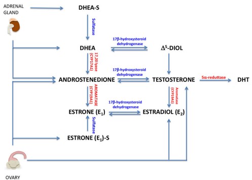 Figure 4. Circulating androgens in women
(Source: Giovannelli P, et al. 2018)
The two main manifestations of polycystic ovary syndrome (PCOS) are hirsutism and anovulatory infertility, although other metabolic disorders include insulin resistance, diabetes and obesity. Hyperandrogenemia clearly suggests PCOS. As we now know, a hyperandrogenic milieu in the womb can cause the epigenetic reprogramming of foetal reproductive tissue to break down and create PCOS in adults. Interestingly, when tested in consistent genetic contexts, elevated androgen levels change the methylation of four human PCOS genes: luteinising hormone/choriogonadotropin receptor (LHCGR), follicle-stimulating hormone receptor (FSHR), insulin receptor (INSR) and DENND1A in oocytes. Additionally, excess androgens are closely linked to the development of metabolic disorders. Androgen apical hypertrophy and increased pro-inflammatory factors release from fat tissue to mediate an inoflammatory low-level inflammatory cascade. In addition, boosted androgens result in selective accumulation of visceral fat in the abdomen and an increase in subcutaneous adipocyte numbers, which increases insulin resistance, LH levels and adrenocorticotropic hormone (ACTH) and metabolic malfunction.
Breast cancer is among the most common malignant tumours that women get, and it can pose an immense health risk to the patient. We know from clinical trials of breast cancer that androgens and the AR have been linked to disease. This AR signalling network not only reveals the aggressiveness of the disease but also promises possible therapy for breast cancer. AR can effectively inhibit the proliferative effects mediated by ER. Some researchers suggest that AR's primary mechanism of action is to control ERα levels by enhancing ERβ levels. ERβ, in turn, regulates the transcription of proliferation-related genes by binding with ERα as a co-transcription factor. There is also an interaction between AR and HER2; upon activation, AR can significantly increase the expression of HER2 mRNA. Studies have shown that there is a positive feedback regulation between AR and HER2 overexpression signaling, where AR activation enhances HER2 expression, subsequently promoting extracellular signal-regulated kinase (ERK) phosphorylation, which then stimulates AR in return.
References
| Target | Cat. No. | Product Name | Size | Species Reactivity | Application | Detection Sample | |
| PARM1 | DEIA-FN1079 | Human PARM1 (Prostate androgen-regulated mucin-like protein 1) ELISA Kit | 96T | Quantitative | Serum, plasma, cell culture supernatants, tissue homogenate | Inquiry | |
| DEIA-FN1080 | Mouse Parm1 ( Prostate androgen-regulated mucin-like protein 1 homolog) ELISA Kit | 96T | Quantitative | Serum, plasma, cell culture supernatants, tissue homogenate | Inquiry | ||
| AR | DEIA-XYA855 | Androgen Receptor ELISA Kit | 96T | Qualitative | Nuclear, cell lysates | Inquiry | |
| DIA-XYA123 | Androgen Receptor ELISA Kit | 96T | Qualitative | Cultured cells | Inquiry | ||
| DEIA3174 | Human AR(Androgen receptor) ELISA Kit | 96T | Human | Quantitative | Serum, plasma, tissue homogenates and other biological fluids. | Inquiry | |
| DEIA-XYA857 | Androgen Receptor (Phospho-Ser650) ELISA Kit | 96T | Qualitative | Nuclear, cell lysates | Inquiry | ||
| DEIA-XYA860 | Androgen Receptor (Phospho-Tyr363) ELISA Kit | 96T | Qualitative | Nuclear, cell lysates | Inquiry | ||
| DEIA-XYA856 | Androgen Receptor (Phospho-Ser650) ELISA Kit | 2 x 96T | Qualitative | Cultured cells | Inquiry | ||
| testosterone | DEIA05747 | Testosterone ELISA Kit | 96T | N/A | Quantitative | Saliva | Inquiry |
| DEIA1609 | Testosterone Human ELISA Kit | 96T | Human | Quantitative | Serum, plasma | Inquiry | |
| DEIA2246 | Testosterone ELISA Kit | 96T | Quantitative | Serum, plasma | Inquiry | ||
| DHT | DEIA1899 | Dihydrotestosterone ELISA Kit | 96T | Human | Quantitative | Serum | Inquiry |
| DEIA-XY62 | Human 5 alpha-Dihydrotestosterone ELISA kit | 96T | Human | Quantitative | Serum | Inquiry | |
| DEIA-NS2307-118 | DHT(Dihydrotestosterone) ELISA Kit | 96T | Universal | Quantitative | Serum, plasma, tissue homogenates and other biological fluids. | Inquiry |
| Target | Cat. No. | Product Name | Expression System | Tag/Conjugate | Application | |
| DHT | DAG2982 | Dihydrotestosterone-3 [BSA] | N/A | BSA | N/A | Inquiry |
| DAG1092 | Dihydrotestosterone [HRP] | N/A | HRP | N/A | Inquiry | |
| Methyldehydrotestosterone | DAGA-168B | Methyldihydrotestosterone [BSA] | N/A | BSA | LFIA | Inquiry |
| DAGA-168K | Methyldihydrotestosterone [KLH] | N/A | KLH | Immunogen | Inquiry | |
| DAGB354 | Methyldehydrotestosterone [HRP] | N/A | HRP | IA | Inquiry | |
| testosterone | DAGA-146K | Dihydrotestosterone [KLH] | N/A | KLH | Immunogen | Inquiry |
| Testosterone | DAGA-181K | 17a Testosterone [KLH] | N/A | KLH | Immunogen | Inquiry |
| DAG-WT2662 | Testosterone control | N/A | Unconjugated | Immunoassays | Inquiry | |
| DAG3024 | Testosterone-19 [BSA] | N/A | BSA | N/A | Inquiry |
Loading ......
