Immunohistochemistry (IHC), based on the principles of immunology, employs a highly specific antigen-antibody reaction to detect target molecules in tissues or cells. It involves extracting antigens or semi-antigens from tissues or cells, generating specific antibodies through immunization, and using these antibodies to identify similar antigenic substances. By employing histochemical methods, such as fluorescein, enzymes, metal ions, or isotopes, the binding sites of antigens and antibodies can be visualized, allowing for qualitative, localization, and quantitative analysis of unknown antigens.
Pathological diagnosis is a diagnosis of disease made by observing the general changes of organs, tissue structure, and cellular lesions under the microscope, so it is more objective and accurate than the analytical diagnosis made based on the clinical history, symptoms, and signs, as well as the diagnosis made by using a variety of images (e.g., ultrasonic waves, X-rays, CTs, MRIs, etc.). In the diagnostic field, pathological diagnosis is usually the final diagnosis of the disease, with the highest authority, and is the "gold standard" in the industry, which can provide clinics with important information such as individualized medication, prognosis assessment, efficacy monitoring, early diagnosis and early screening.
The field of pathological diagnosis can be divided into four major areas, including histopathology, cytopathology, immunohistochemical pathology, and molecular pathology. Among them, IHC is widely used in pathological diagnosis, which can provide objective evidence at the level of protein expression and help pathologists accurately determine the nature and characteristics of diseases, especially in tumor diagnosis. With the invention of monoclonal antibodies, peroxidase/anti-peroxidase (PAP) method, avidin-biotin complex method (ABC method), thermal antigen retrieval, and other technologies, the immunohistochemical signal amplification detection system has made great progress.
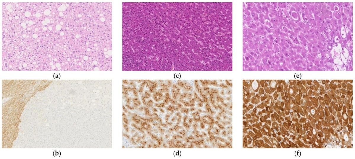 Fig. 1 Histological appearance and results of immunohistochemical staining of hepatocellular adenoma (HCA). (Takahashi Y, et al., 2021)
IHC is widely used in pathological diagnosis, including judging benign and malignant tumors, determining the source of tumor cells, differential diagnosis of tumor types or subtypes, tumor grading, prognosis judgment, targeted therapy, and the discovery and determination of micrometastases. In addition, immunohistochemistry can also be used for the determination of tumor targets for targeted drugs, the prediction of response to tumor chemotherapeutic drugs, and the comprehensive evaluation of tumor prognosis.
Distinguishing benign and malignant tumors
IHC has proven invaluable in distinguishing between benign and malignant tumors. By utilizing immunoglobulin (Ig) light chain antibodies, the monoclonal or polyclonal nature of B lymphocyte proliferation can be detected, aiding in the differentiation of reactive hyperplasia from neoplastic hyperplasia.
Tumor staging and prognostic assessment
Accurate tumor staging is crucial for determining prognosis and selecting appropriate treatment strategies. IHC plays a vital role in detecting critical indicators, such as infiltration, lymphatic or vascular invasion, and molecular markers. By employing IHC, clinicians can assess these changes, enabling precise tumor staging and providing valuable prognostic information.
Identification of cell properties and tumor origins
IHC enables the identification of specific antigen components within cells, facilitating the determination of cell properties and tumor origins.
Diagnosing metastatic tumors of unknown origin
Metastatic tumors of unknown origin pose significant diagnostic challenges. However, IHC can aid in determining the histological source of malignant tumors and the primary site. By leveraging specific antibodies, such as the keratin antibody (CK20) for gastrointestinal cancers, bile duct cancers, and pancreatic cancers, we can accurately identify the primary tumor site. This information is critical for guiding treatment decisions and improving patient outcomes.
Classifying "undifferentiated" malignant tumors
Undifferentiated malignant tumors, including cancers or sarcomas, present a diagnostic dilemma due to their lack of differentiation markers. IHC offers a powerful tool for their classification by detecting specific antigens associated with particular tumor types.
Detection of micrometastasis
Conventional tissue sections often pose challenges in identifying individual or small clusters of metastatic tumor cells. Additionally, distinguishing sinus histiocytosis in lymph nodes from early metastasis of certain cancers can be problematic. However, IHC provides a solution by enabling the timely and accurate detection of minute cancer metastases.
Diagnosis of infectious diseases
IHC offers a powerful diagnostic tool by utilizing specific antibodies against viral, bacterial, fungal, and parasite antigens to detect and diagnose various infectious diseases. Through IHC, pathogenic microorganisms like hepatitis B virus (HBV), human papillomavirus (HPV), herpes virus (HSV), and cytomegalovirus (CMV) can be identified accurately. The use of IHC in infectious disease diagnosis provides several advantages, including timely detection, high sensitivity, specificity, low risk, and overall effectiveness.
IHC is an indispensable tool in the field of pathological diagnosis, providing valuable insights into disease understanding, tumor classification, and prognostic assessment. As a leading provider of innovative solutions, Creative Diagnostics offers a comprehensive range of high-quality antibodies, including Her2, PD-1, Ki-67, Tim3, CTLA4, Lp-PLA2, Muc2, Cytokeratin 18, BRAF V600E, IDH1 R132H, MLH1, MSH2, MSH6, and PMS2 for IHC applications, enabling precise and reliable results in pathological diagnosis. With our extensive portfolio and expertise, we empower clinicians and researchers to unravel the complexities of diseases and make informed decisions for improved patient outcomes.
Reference