
Inquiry Basket
 Loading ......
A mono-chain, simple hormone produced by parathyroid gland chief cells is called PTH, meaning parathyroid hormone. It tends to regulate the body's calcium and phosphorus metabolism mostly by causing blood calcium to increase and blood phosphorus to decrease. Sometimes these diseases – parathyroid adenoma, hyperplasia and carcinoma – produce too much PTH and cause metabolic diseases. In chronic renal failure patients on long-term dialysis, these imbalances of vitamin and mineral metabolism can result in decreased blood calcium and higher blood phosphorus levels that stimulate the parathyroid glands to produce and secrete more PTH.
PTH is generated in the parathyroid glands and diffused into the blood. The entire PTH is a linear polypeptide composed of 84 amino acids, and weighs in at about 9500. The life-activity relies on the 27 first amino acid residues at the N-terminus. It has a few minutes of half-life for the biologically active N-terminal section. We can directly measure parathyroid secretion with selective measuring of total PTH.
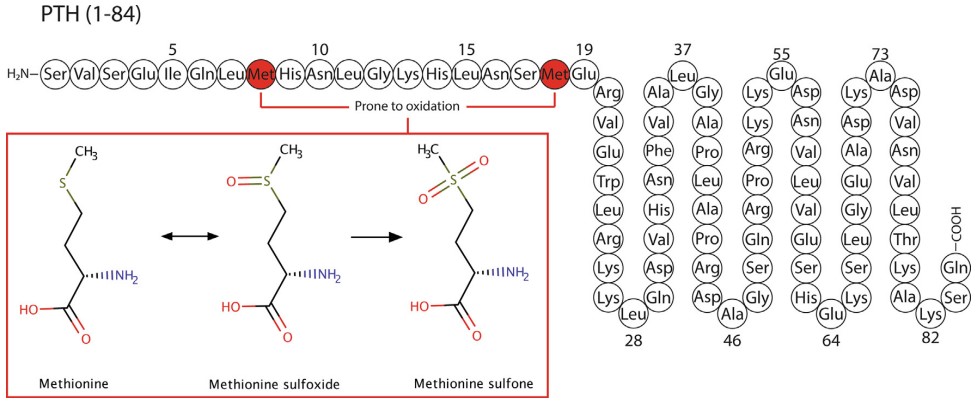 Figure 1. PTH consists of 84 amino residues
(Source: Ursem SR, et al. 2020)
In the blood, PTH exists in three forms:
1. Complete PTH or PTH (1-84): it is biologically active, reaching 5–20% of total PTH. It has a very, very short half-life, around 2–4 minutes.
2. Carboxy-terminal PTH segments: it is inactive. These are 75%-85% of all PTH like PTH (36-84); PTH (44-84) and PTH (53-84). They have 25-60 minutes half life.
3. Amino-terminal PTH fragments, that are biologically active, like PTH (1-34). They are in lower blood levels and have a short half-life.
The physiological effects of PTH include:
1.Bone effects: PTH activates osteoclasts, reprogrammes osteoclasts, and temporarily turns off osteoblast activity. This helps the bone to resorb and dissolve faster and the calcium is displaced from the bone and sucked out into the bloodstream boosting blood calcium levels.
2.Affect Kidneys: PTH makes phosphate not reabsorbed in the proximal renal tubules thus release more phosphate into the urine. The consequence of this is that too much PTH leads to hypophosphatemia and hyperphosphaturia. By contrast, PTH reuptakes calcium from the distant kidney tubules.
3.Recrease in Renal 1-α-hydroxylase Activity: PTH increases the synthesis of calcitriol (active vitamin D) in the kidneys which in turn improves calcium absorption in the gut.
4.Autoregulation of Cell Functions: PTH is the regulator of various cells functions such as cardiomyocytes, adipocytes and pancreatic β-cells.
OP decreases bone mass and density through the disassembly of bone-growth to bone-decomposition. It is Osteoporotic Disease, the most common metabolic bone disease. PTH manages the remodeling of bone by binding to its receptor (PTH1R). When the PTH stimulation is repeated daily then more bone will be reabsorbed and when it is intermittent then more bone will be produced.
BMSCs (bone marrow mesenchymal stem cells) are initially transformed into osteoprogenitor cells, then pre-osteoblasts, and finally mature to mature osteoblasts (OB). OB then go to bone lacunae to release bone matrix that's then mineralised to create new bone. During this process, intermittent stimulation by PTH promotes the proliferation of pre-osteoblasts, increases the number of OB, and ultimately facilitates osteoblastic differentiation.
Once PTH has binded to PTH1R, bone metabolism-related signalling pathways control osteoblastic differentiation, dependent on the amount and type of stimulation. It's cAMP/PKA pathway, PLC/PKC pathway, non-PLC/PKC pathway, and Wnt/β-catenin pathway that play a role. PTH also changes the cAMP/PKA signaling pathway, which controls both bone formation and resorption. The PTH was found to co-transcribe the co-transcription factor CITED1 into and out of the nucleus via this signalling network, controlling gene expression for genes such as osteocalcin (OC) and bone alkaline phosphatase (BALP), which is involved in bone metabolism. Periodic PTH stimulation enables differentiation of BMSCs into osteoblasts through regulation of the PLC/PKC signalling system and up-regulation of osteogenic genes like Runx2 and BALP.
 Figure 2. Parathyroid hormone (PTH) and bone metabolism
(Source: Evenepoel P, et al. 2016)
PTH is primarily used to maintain homeostatic calcium (important for the cardiovascular system). A high circulation PTH level is inversely associated with left ventricular hypertrophy and is also inversely associated with NT-proBNP levels in heart failure and ejection fraction. Increased PTH can attach to receptors on heart failure patients' smooth muscle cells and activate elevated levels of oxidative stress, endothelial dysfunction and intracellular overload of calcium through adrenergic receptors and L-type calcium channels resulting in heart failure as a vicious cycle.
Research has demonstrated that PTH can stimulate cardiac influx of calcium ions, and so release norepinephrine, increasing the contractility of the myocardium and ultimately leading to cardiac hypertrophy. Heart muscle cells – the heart's healthy cardiomyocytes – also have L- and T-type calcium channels, and the closure of the L-type channel protects the cell from oxidative stress. High levels of PTH in the blood can open G protein-coupled L-type calcium channels in cardiomyocytes, increasing calcium ions that get into the cells. PTH1R and PTH-related proteins can also stimulate PTH-associated pathways in excess releasing intracellular calcium. Overloading with calcium interrupts normal mitochondrial ATP production, oxidative stress, and is the pathological hallmark of myocardial necrosis. PTH may also exacerbate the process of replacement of myocardial necrotic tissue by fibrous tissue. Myocardial fibrosis was found to be aggravated in rats with left ventricular hypertrophy after PTH intervention. This result may at least partially indicate that high PTH levels aggravate myocardial fibrosis.
The RAAS is an important fluid-control system in our body. This system includes renin, angiotensinogen, angiotensin I, angiotensin-converting enzyme, angiotensin II, angiotensin II type 1 receptor and aldosterone. RAAS modulates the water and salt metabolism. Chronic activation of RAAS is a contributing factor to the onset and persistence of heart failure. PTH can influence the secretion of RAAS through various pathways. PTH can promote release of norepinephrine from the atria and kidneys by activating PTH1R. The renal sympathetic nerve is stimulated, which lead to increase renin secretion and activate RAAS. Short- and long-term use of angiotensin-converting enzyme inhibitors can reduce serum PTH, indicating an interaction between PTH and RAAS.
 Figure 3. Direct and indirect effects of PTH on the cardiovascular system
(Source: Fujii H. 2018)
Secondary hyperparathyroidism or SHPT is among the most frequent and severe complications of chronic renal failure. SHPT can cause multi-system disorders in patients, including skin, bone, cardiovascular system. SHPT can be found throughout the course of chronic kidney disease (CKD), but its pathogenesis is quite complex. Among these, decreased blood calcium, increased blood phosphorus, and increased PTH levels are hematological indicators of prevalent abnormalities in SHPT.
The calcium-sensing receptor or CaSR is a coupled protein located on parathyroid cell membranes. It has a structure characteristic of the G protein-coupled receptor family. The extracellular domain contains many highly specific calcium-binding sites, when binding with calcium ions, it will be activated. This activation triggers intracellular signaling pathways that ultimately lead to rapid inhibition of PTH secretion. After CaSR activation, as blood calcium levels increase, PTH levels decrease. However, there are fewer CaSRs and less concentrations of calcium ion with progressive loss of renal function. It will lead to release more intracellular PTH, and increase more PTH into the blood.
Due to the different forms and biological characteristics of PTH in the blood, clinical PTH testing is categorized into three generations. The first generation primarily used radioimmunoassay methods for PTH detection, but it has been phased out due to limitations in sensitivity and accuracy. The second generation of testing measures PTH, including PTH (1-84) and PTH (7-84), which is commonly referred to as PTH testing or iPTH testing in clinical practice. The third generation of testing specifically measures biologically active PTH (1-84), which is what is clinically referred to as PTH (1-84) testing.
PTH levels are one of the important metrics in parathyroid surgery success, and PTH management can offer helpful guidance for future clinical treatment of patients with chronic kidney disease. Primary hyperparathyroidism or PHPT: Excess PTH is released by parathyroid adenoma, hyperplasia, carcinoma or cysts and causes systemic abnormalities in calcium and phosphorus metabolism. There is no cure other than surgical removal. Measurement of PTH at parathyroid adenoma excision, it's suggested, can also predict successful surgery. It can determine if the parathyroid adenoma has been completely removed, and if all the hyper-functional parathyroid tissue is removed.
SHPT is a chronic compensatory clinical manifestation, which is characterized by parathyroid hyperplasia and excessive PTH synthesis and secretion. PTH measurement in surgery can help doctors to remove glands, and improve the success rate of the surgery. Some researchers believe that different PTH fragments decrease at different rates. Moreover, the metabolic pattern of PTH is initially rapid followed by a slower decline; therefore, it is insufficient to evaluate surgical success solely based on whether PTH levels return to normal within 10 minutes after resection. A failure to normalize PTH levels within this timeframe may be related to continued stimulation of the glands during intraoperative exploration.
References
| Target | Cat. No. | Product Name | Size | Species | Application | Detection Sample | |
| PTH | ABPR-ZB224 | Human Parathyroid Hormone/PTH Antibody Pair Set | 5 Plates, 15 Plates | Human | sELISA | Inquiry | |
| ABPR-0738 | Human PTH ELISA Matched Antibody Pair | ELISA | Inquiry | ||||
| DEIA1826B | Bovine Intact Parathormone ELISA Kit | 96T | Quantitative | Serum, plasma, tissue homogenates and other biological fluids. | Inquiry | ||
| DEIA1836 | Human Intact-PTH ELISA Kit | 96T | Human | Quantitative | Human Serum | Inquiry | |
| DEIA-BJ659 | Mouse I-PTH(intact PaRathormone) ELISA Kit | 96T | Mouse | Quantitative | Serum, plasma, tissue homogenates, other biological fluids | Inquiry | |
| DEIABL239D | PTH (1-84) Depletion Kit | 10 x 96 tests | N/A | Qualitative | Serum; Plasma. | Inquiry | |
| DEIABL239 | PTH (1-34)-Teriparatide ELISA Kit | 96T | Human | Quantitative | Serum | Inquiry |
| Target | Cat. No. | Product Name | Expression System | Tag/Conjugate | Application | |
| PTH | DAG-WT2730 | Human Parathyroid Hormone (PTH) (1-34) peptide | N/A | N/A | N/A | Inquiry |
| DAG-WT2731 | Human Parathyroid Hormone (PTH) (39-84) peptide | N/A | N/A | N/A | Inquiry | |
| DAG-WT1941 | Recombinant Human PTH Protein [GST] | E. coli | GST | WB, ELISA | Inquiry | |
| DAG-WT1942 | Recombinant Human PTH Protein [hFc] | HEK293 cells | hFc | WB, ELISA | Inquiry | |
| DAG-IV03 | Recombinant Human PTH Protein | E.coli | N/A | Inquiry |
Loading ......
