Introduction of Flow Cytometric Analysis of Microparticles
Extracellular vesicles are small buds of the cell membrane about 40–1,000 nm in diameter. Currently, three types of extracellular vesicles have been defined: microparticles (MPs) generally range from 100 to 200 nm in diameter but can be up to 1,000 nm in size; exosomes (EXs) range from 40 to 100 nm; and apoptotic bodies (AB) vary considerably in size and are usually large (>1.5 mm). There are differences in the mechanisms of formation between the vesicle types, which likely affect their function. Although it is clear that MP formation is an active process which is continually being performed by cells and enhanced by agonists, the precise molecular mechanisms remain to be determined. The formation of MPs does depend on the reorganization of cytoskeleton. MPs contain a variety of intracellular components, including cytoplasmic proteins, RNA, and DNA, and generally express the proteins from the parent cells. EXs are derived from multivesicular bodies and consequently contain only cytoplasmic proteins. Apoptotic body formation is a terminal step in apoptosis where the cell has released MPs beforehand, activated caspases, fragmented DNA, and dissociated into smaller particles, presumably to facilitate phagocytosis.
Initially, extracellular vesicles were considered cellular debris but accumulating evidence indicates that they play important functions in homeostasis. Dysregulation of MP production, in particular, is a biomarker for disease and these MPs may be a required pathway leading to pathogenesis. MPs are ubiquitous being formed from every cell type, including nonnucleated platelets and erythrocytes. Activation of platelets by thrombin, ADP, and other agonists leads to the translocation of phosphatidylserine from the inner leaflet to the outer leaflet and the adoption of a high affinity conformation by integrins, such as CD41/ CD61 (also known as aIIb/b3). The negatively charged surface caused by increased levels of phosphatidylserine in the outer leaflet is important for the binding of the prothrombinase and tenase complexes. The release of MPs from activated platelets markedly increases the surface area for the binding of the tenase and prothrombinase and facilitates the activation of the coagulation cascade. The importance of MPs in the activation of the coagulation system is underscored by the ability of MPs to markedly decrease coagulation times. In nucleated cells, agonists that elicit MP formation (e.g., lipopolysaccharide) often also elicit apoptosis. We have reported that MPs can carry functional molecules from the membrane of one cell to another, providing an important new signaling mechanism that falls outside of the conventional autocrine or paracrine signaling by secreted molecules. Increased numbers of circulating MPs have been found as biomarkers in a number of diseases (cerebral malaria, sepsis, and several autoimmune diseases), and our data indicates that they may play a required role in the pathogenesis of experimental cerebral malaria. Antigen-presenting cells of the immune system are an important functional source of EXs. EXs exhibit marked adjuvant effects, improving the immune response to vaccine candidates and contain functional MHC II and peptide along with costimulatory molecules. Apoptotic bodies are considered inert and not to elicit a response by immune cells. Cell-derived vesicles are a new mode of cell–cell signaling that is now under intense investigation. MPs in infectious disease may also exhibit other important functions, such as carrying antigens that are important for the activation of the immune system.
To determine the molecular mechanisms of MP formation, their function and production from selected cell types in response to agonists, it is necessary to separate out the individual cell populations. The process of separating vesicles from cells and soluble factors by centrifugation. The resultant MPs or MPs produced in blood must be first separated from their parent cells and soluble factors so that these variables do not affect the functional analysis of MPs. This step is critical especially if the MPs are frozen for subsequent analysis.
Methods of Flow Cytometric Analysis of Microparticles
Collection and Processing of Blood for MP Analysis
- Human blood. The assessment of the number and phenotype of blood MPs during disease requires the standardization of collection and processing, as well as the minimization of steps that may actually elicit MP formation from blood cells. Steps that should be standardized for the analysis of patient samples include: sampling site, the size the needle (21G) used during venipuncture, the absence of a tourniquet, the anticoagulant, and the use of a 10 mL vacutainer rather than a syringe (constant suction). Apyrase (1 U/mL) and either PGE1 (100 ng/mL) or PGI2 (0.2 mM) may be added to further inhibit platelet activation, but we have found that this is not necessary. Donors should not have taken medication (especially one interfering with platelet functions, such as aspirin) for 10 days prior to blood sampling. The samples should not be shaken, chilled, or overheated because shear and temperature stress induces increased MP formation. Once collected, the blood should be rapidly processed within 1 h to prevent further release of MPs from cells. If platelet MPs are a component of the analysis, 1 mL of blood is removed for flow cytometry to provide a positive control for platelet labeling and analysis. To directly analyze MPs in blood, it is necessary to generate cell-free enriched MPs via a two-step centrifugation procedure.
- Centrifuge the blood at 1,500×g for 15 min to generate about 4.5 mL of platelet-poor plasma.
- Carefully remove the supernatant and pipette 1.5 mL into microcentrifuge tubes. Centrifuge at 13,000×g for 2 min to remove all residual platelets and cell fragments larger than 1 mm. The supernatant is MP enriched, platelet-deficient plasma or "MP plasma."
- Platelets are the smallest cell type in the blood, so the quality of cell-free, MP-enriched plasma ("MP plasma") is assessed by counting the number of CD41+ platelets present in 1 mL of the blood versus 1 mL of MP plasma. If the MP plasma cannot be analyzed immediately on site, the MP plasma can be frozen at −80°C and analyzed at a later time (see Note 1). Because the cellular sources of MPs have been depleted, this procedure does not, in our experience, change the MP counts significantly.
- This MP plasma does contain EXs. If it is important to distinguish between the two vesicle populations, then centrifuge the MP plasma at 10,000×g for 1 h to pellet the MP; the majority of the EXs remain in the supernatant. Purification of the EXs requires centrifugation on a sucrose gradient. Note, some investigators centrifuge at 20,000×g, which pellets vesicles of >200 nm size; we have not, however, tested this possibility. The pelleting of MPs is also used to separate soluble factors (e.g., cytokines) in the blood from the MPs; this is necessary to ascertain the function of MPs. The MPs primarily pellet and EXs remain in solution; this separation is, of course, not absolute but does provide a mechanism to analyze each vesicle population.
- Mouse blood. Collect blood samples via bleeding from the retro-orbital plexus or intracardiac puncture, and collect the blood (1:4) into citrate. In our hands, bleeding from the retro-orbital plexus yields few if any activated platelets, and no evidence of coagulation (assessed by coagulation factors in the blood). Process the blood into MP plasma as described in step 1 (see Notes 1 and 2).
- Rat blood. Obtain blood by cardiac puncture or from an indwelling catheter.
Generation of Microparticles from Cells
MP can be generated from a mixed population of cells, e.g., whole blood or from purified cells (cell culture, isolated subpopulation). If the purpose is to increase the number of MP present in the solution, an agonist that will induce the release of MP without modifying the cell surface phenotype, i.e., no up- or downregulation of adhesion molecules on endothelial cells or glycoprotein on platelets, can be chosen, e.g., calcium ionophores: calcimycin and ionomycin. Because all cell types release a basal level of MP, the cell supernatant always contains some MPs and their number is increase in a time dependently manner regardless of the presence of an agonist. These MPs are used as "control" MP. The biological questions being addressed determine which cell-specific and activation markers are included in the flow cytometry analysis. Below, we briefly summarize protocols for generating in vitro MPs from these selected cell types (see Note 3).
- Platelets.
- Centrifuge the blood at 100×g for 10 min to pellet erythrocytes and leukocytes and consequently generate Platelet Rich Plasma (PRP).
- Generate purified platelets by washing PRP with Tyrode's buffer lacking Ca and Mg or by Sepharose CL-4B gel filtration. Wash platelets by centrifuging at 12,000×g for 1 min.
- Enumerate the platelets in a hemocytometer, e.g., an Improved Neubauer chamber, or by flow cytometry using counting beads or by using a cytometer that determines the number of particles per unit volume. Resuspend the platelets at 1×10⁷ /mL in 0.1 mL aliquots and then stimulate with the agonists for the indicated times (see Note 4).
- Erythrocytes. Erythrocytes are the predominant cell type in blood and are easily purified from other cell populations.
- Centrifuge the blood at 120×g for 10 min; remove both the supernatant and buffy coat (thin layer of white cells on the surface of the erythrocyte pellet) in order to generate purified erythrocytes.
- Centrifuge the blood twice at 120×g for 10 min, each time removing the supernatant and any residual buffy coat.
- Ascertain the purity of the erythrocyte population by flow cytometry using anti-CD41 to label platelets, anti-CD45 to label leukocytes, and anti-CD235a (glycophorin A) to label the erythrocytes. Addition of counting beads determines the numbers of RBCs/mL.
- Count the RBCs, resuspend at 1×10⁷ /mL in 0.1 mL aliquots, and then stimulate with the selected agonists.
- Leukocytes. Purify primary mononuclear cells from blood by slowly layering the blood on top of Histopaque-1077, centrifuging (400×g for 30 min at room temperature), and collecting the mononuclear cells at the interface. Further separate the individual cell populations either by Fluorescence Activated Cell Sorting (FACS) or Magnetic Activated Cell Sorting (MACS) using the cell population specific antibodies. Stimulate the purified cell populations with the selected agonists in vitro. Centrifuge the cells to separate the cells from MPs for flow cytometry. A variety of cell lines are available for the analysis of MP formation in vitro, which does not require cell purification as described above. However, their transformation into immortal cells may alter their responses to agonists and MP generation. These cells are cultured until in the log phase of replication and then used in the assays.
- Endothelial cells. Culture primary or cell lines on a matrix, such as collagen or gelatin. When reaching subconfluence, cells are stimulated during various periods of time (from 10 min with histamine to 18 h with TNF). Because endothelial cells are adherent, the MP are detected in the culture supernatant without purification step. However, an enrichment/concentration by centrifugation may be wanted to identify poorly represented subsets of MP, i.e., MP carrying low levels of antigens or antigens localized at certain sites on the cell surface and therefore not present on all MP.
For in vitro analyses of MP formation, the membranes of the cells can be fluorescence labeled, confirming the origin of the MPs. Cells are labeled green with CFSE or PKH67 and red with PKH26. CFSE is a colorless "vital" dye passively diffuses into cells until it is cleaved by intracellular esterases removing the acetate group, thereby becoming brightly fluorescent. The succinimidyl ester group reacts with intracellular amines, thereby forming fluorescence that is retained within the cells. The cells are incubated in serum-free medium for 15 min at 37°C (platelets can be labeled at room temperature) in 10 mM CFSE (range: 0.5–10 mM) or using the 2× dye solutions and diluents for the PKH dyes. The cells are washed to remove excess dye and then used in the assay. Pelleted MP can be labeled directly with the same dyes, and Wagner's group has also labeled MPs with Calcein AM (5 mg/ mL) for 20 min at room temperature.
Fluorescence Labeling of Microparticles
If the MP plasma was frozen, thaw the samples rapidly using a water bath and then place it on ice. As described above, activated cells exhibit increased intracellular Ca2+ that in turn induces translocation of phosphatidylserine from the inner leaflet to the outer leaflet; thus, most investigators use annexin V labeling to identify MPs. However, others indicate that the phosphatidylserine may not be accessible or expressed on all the MPs (particularly of endothelial cell origin), resulting in an undercounting of MPs.
- Platelets: all platelets express CD41/CD61 (gpIIb/IIIa) on their surface and CD41 is only detected on the cell surface of platelets. Accordingly, anti-CD41 is used to identify platelet-derived MPs (PMPs). The activated conformation of CD41/ CD61 is recognized by PAC1 (humans) and JonA-PE (mouse). The von Willebrand Factor receptor CD42b-CD42c/CD42a/ CD42d (gpIb-ab/gpV/gpIX) is another important platelet protein complex, which also binds thrombin and P-selectin. CD42b is an activation marker that is proteolytically cleaved upon platelet activation, whereas CD42c is constitutive and represents an alternate platelet marker to CD41. P-selectin is detected on the surface of activated platelets. CD41+ and CD42+ MPs may be elicited differentially during pathogenesis, so enumeration of both populations may be indicated.
- Erythrocytes: CD235a (glycophorin A) is used to identify red blood cell-derived MPs (RMPs).
- Leukocytes: CD45 is present on all leukocytes and is used to identify leukocyte-derived MPs (LMPs). For leukocyte subsets, the following mAbs are used: CD14/CD11b (monocytes), CD66b (granulocytes), CD4 (TH), CD8 (TC), and CD20 (B cells) (see Note 5). Because CD14 is a raft-associated protein, Freyssinet and colleagues suggest that CD14 may not be present on MPs or at lower levels; others, however, have used this marker.
- Endothelial cells: CD105 (endoglin) is not only expressed on ECs, but also weakly expressed on leukocytes. Endothelial-derived MPs (EMPs) are therefore: CD105+, CD45−. CD144 (VE-cadherin) is also used as marker for EMPs. CD54 (ICAM1), CD62E (E-selectin), CD62P (P-selectin), and CD106 (VCAM1) can be markedly upregulated on endothelial cells by various agonists and therefore may provide a measure of whether subsets of EMPs are derived from activated endothelial cells.
Fluorescence Labeling of the MPs for Flow Cytometric Analysis (no annexin V Labeling)
- Pipette 10 mL aliquots of MP plasma into flow cytometry tubes.
- Some investigators add 1 mL of EDTA (25 mM) to MP plasma (final concentration 2.5 mM EDTA). EDTA reduces nonspecific labeling but in our opinion is not necessary. It may also affect the binding to some molecules, such as CD41.
- Add 2 mL of selected cell-specific mAbs (final concentration: 15 mg/mL) and incubate for additional 25 min.
- Add 500 mL of HBSS and subject to flow cytometry (see Note 6).
Fluorescence Labeling of the MPs for Flow Cytometric Analysis (with annexin V Labeling)
- 10 mL aliquots of MP plasma are pipetted into flow cytometry tubes.
- Add 1 mL of ready-to-use annexin V-FITC+1 mL of binding buffer 10× (to optimize the calcium concentration necessary for annexin V binding in the plasma and counteract the binding inhibitory effect of the anticoagulant); incubate for 10 min. However, one must be aware that the presence of binding buffer containing 2.5 mM Ca2+ can trigger plasma coagulation, therefore the total incubation time must be constant and samples must be checked prior to flow analysis for the presence of microclots.
- Add 2 mL of selected cell-specific mAbs (final concentration: 15 mg/mL) and incubate for additional 20 min.
- Dilute samples in 0.5 mL of annexin V binding buffer and analyze by flow cytometry.
The choice of fluorescence label(s) depends on the cell population being analyzed in vitro or if determining the cellular origin of the MPs is important. If the cell membrane has been labeled, vesicles derived from the cell membrane will be labeled. Alternatively, the small vesicles can be labeled with Calcein AM. If secondary reagents are used, then the MPs are pelleted by high speed centrifugation (25,000×g, 30 min, 15°C) before the fluorescence labeled secondary mAb or streptavidin is added. It is important to include negative isotype controls for the selected specific Abs because there is greater nonspecific binding of Abs to MPs than intact cells and the AB may not be removed if washing is omitted.
Polystyrene beads of defined size are often added to the sample to provide rudimentary quality control. In most cases, 1 mm polystyrene beads are added and events exhibiting lower forward scatter are collected and defined as MPs. Polystyrene beads with defined sizes (e.g., 190, 530, 780, and 990 nm) have also been used to provide a guide for size. However, it is important to emphasize that the forward scatter signal derived from these beads cannot be related to the size of the particle. If a known number of beads are added, these can then be used as counting beads if the flow cytometer does not assess the volume of sample analyzed.
Conventional Flow Cytometric Analysis of Microparticles
The analysis of MPs on a Beckman-Coulter's Cytomics FC 500 flow cytometer is summarized. All parameters, including FSC and SSC, are set in log-mode. The parent cell may be used to set the compensation if several fluorophores are used in the analysis. It is important to ensure that saturating levels of antibody are used for fluorescence labeling of the MPs; we recommend performing titration curves using MPs or the parent cells. The MP sample is then acquired on the flow cytometer.
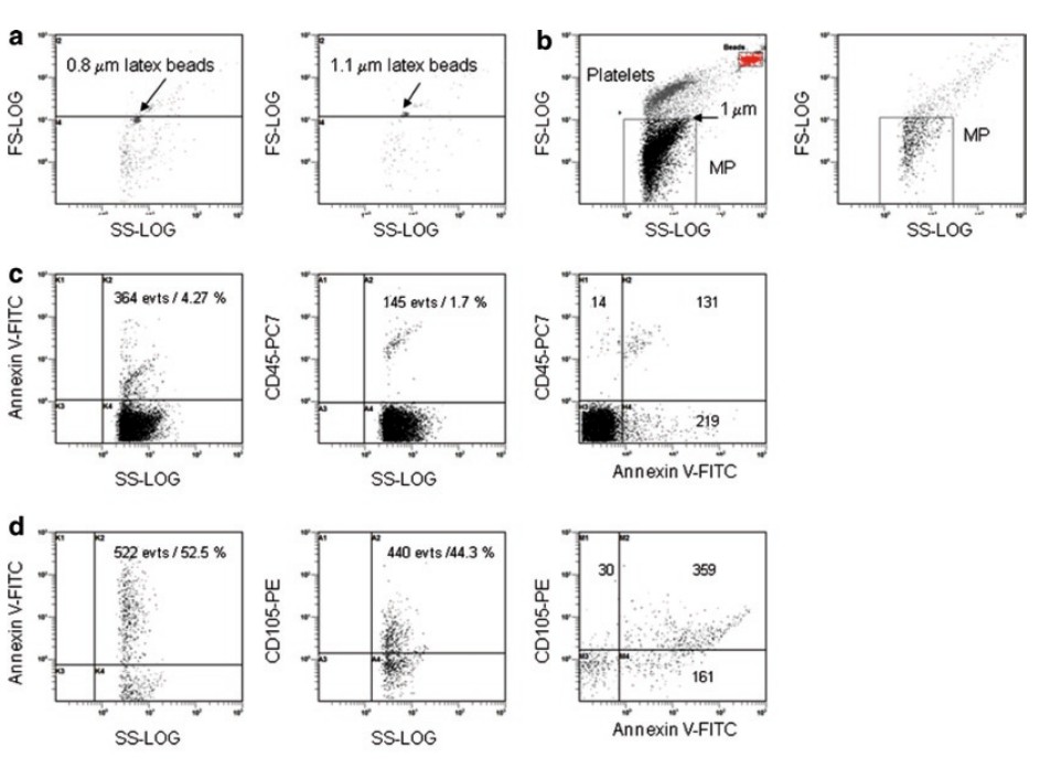 Figure 1. Conventional flow cytometric analysis of MPs.
- Run 0.8 and 1.1 mm particles latex beads alone, and use to set the upper limit of the population to analyze with FSC<1 mm and have these events shown in the fluorescence dot plots.
- Run positive samples (PRP) if applicable to define the platelet region. Note, platelets are the smallest cell type and are about 2–5 mm in size and must be excluded from the MP analysis. Other cell populations are much larger, so they are not problematic.
- To set the positive gate limit, acquire MP events using isotype controls for the specific antibodies.
- To verify compensation, acquire MP events with single specific antibodies.
- Acquire multiple-labeled MP events. The slowest flow rate of the instrument provides the greatest sensitivity because the fluorescence signal is integrated the longest but may take too long to sample. Increasing the flow rate above 300/s will increase the number of coincident events and lower the count assessed by the flow cytometer (see Note 7). When possible, more than 1,000 positive MPs should be collected in each sample, and we save all events for the subsequent analysis.
- Determine the percent positive events and the number of labeled MPs. If the flow cytometer is syringe-driven, then the number of MPs/mL sample is calculated by the instrument. Alternatively counting beads are spiked into the sample. The number of MPs per mL=[number assessed by flow cytometer×(beads counted/beads added)]/sample volume.
A gate is set to include only events exhibiting FSC<1 mm beads; if the MPs are fluorescence labeled, then a second gate is set to include only events with a fluorescence intensity above that observed with unlabeled MPs. Because MPs from many cell populations express PS, fluorescence labeling with annexin V can also be used to positively identify MPs in the sample. The fluorescence intensity is then measured for each MP, which then indicates the cellular origin of the MP and extended analyses may determine functional proteins on the surface of MPs. For example, we have reported that MPs contain the P-glycoprotein (that confers multidrug resistance to cancer cells), bind to drug sensitive cancer cells, and functionally transfer drug resistance. The numbers of positive MPs for a specific protein is also recorded.
Imaging Flow Cytometric Analysis of Microparticles
Amnis has developed an imaging flow cytometer, which overcomes many of the problems associated with flow cytometric assessment of MPs. First, the instrument provides an actual measurement of size rather than forward scatter. Accordingly, it is not necessary to extrapolate the arbitrary forward scatter measurement of polystyrene beads to the size of biological vesicles, which is not valid anyway. Second, the signal to noise ratio for small particles is markedly better by using imaging cytometry. A conventional flow cytometer measures accurately the fluorescence intensity in a selected range of wavelengths of light (bandpass-filtered), but the small particle occupies only a small portion of that volume. Thus, the small signal is present in a large background, which compromises sensitivity. In contrast, the ImageStream analysis software (IDEAS) measures signal specifically with the pixels that the particle's image occupies. Because signal arising from small objects occupy a minimal number of pixels, there is very little pixelassociated noise, and thus the ImageStream is very sensitive to MP fluorescence. This is especially important for the analysis of MPs, which exhibit low levels of markers on their surface and hence low fluorescence intensity when compared with the parent cell. Furthermore, obvious coincident events can be flagged and potentially included in the analysis. If two particles are captured and are separated spatially in the image, then these can be assessed as distinct MPs in the analysis. The MP plasma is fluorescence labeled and then subjected to Imaging Analysis as described below.
- Set up the instrument with an upper limit for the raw maximal pixel intensity (background subtracted intensity of the brightest pixel in the image) set to 100 to eliminate the collection of bead calibration reagent.
- Acquire the unlabeled samples and single color isotype control samples to set up software compensation for the MP analysis. The samples may need to be diluted if there are large numbers of coincident events. We dilute the samples tenfold into a sample volume of 50 mL.
- Acquire the PRP control samples if included.
- Acquire all events except bead calibration reagent in the test samples with at least 5,000 events. Use compensation settings from the single color samples.
- In the analysis phase, set the MP gate to analyze events < 1 mm and platelet gate for events > 1 mm. Depending on the selected fluorescence labels, display the dot plots and place regions to define subpopulations of MPs. The analysis even indicates the number of unlabeled MPs, which exhibit an SSC above background (not shown). Because our sample contained both platelets and MPs, we can compare both populations. The analysis of the regions reports the fluorescence intensity and number of particles. This analysis shows that thrombin activation increased the number of platelet MPs, which decreased their expression of CD42b, a proteolytically cleaved activation marker. The limitation of the ImageStream is the accurate measurement of size for small particles. Particle cross-section area is measured on the ImageStream by placing a mask or limit around the bright field object identified, counting the number of pixels in that region, and then converting the number of pixels to actual size based on physical pixel size of the camera and objective magnification. In the experiment, the objective was 40×, resulting in 0.5 mm2 pixels. The accuracy of this measurement depends on the ability of the software to place the mask and the focal position of the object. Objects that have low contrast or are smaller than 0.5 mm2 are under masked and appear smaller. If the object drifts from the optimal focal plan, its image blurs and the software will mask this event as larger than its actual size. The ImageStream therefore has limitations on the assessment of MP size, but we contend that this size discrimination is not possible by conventional flow cytometry either.
Alternative Bulk Assays for the Analysis of Microparticles
- An alternate approach to assess MP levels is ELISA MP capture assays; in this ELISA, the MPs are captured by annexin V or antibodies specific to antigens on surface of MPs (e.g., CD41). The levels of MP antigen are then assessed by using a second MP-specific antibody with a colorimetric, fluorescence, or luminescence detection system. This approach while quantitative does not determine the number or phenotype of particles and is sensitive to cell carryover, which gives a large signal compared with MPs.
- Scanning electron microscopy is used to detect the presence of MPs in samples and is the most accurate approach to generate size ranges for the MPs. Confocal microscopy is useful for detecting the binding and potential transfer of functional proteins from MPs to cells. The flow cytometric assays should be extended by performing functional assays to link increases in the selected phenotype of MPs with a function.

 Loading ......
