The development of a test for anti-doping control of erythropoietin(EPO), a hormone used in endurance sport to stimulate red blood cell production, has been a long and exacting task. The method is based on differentiation of natural and recombinant(used in case of doping) hormones in urine by their isoelectric profiles. For this, urine is first submitted to ultra-filtration to concentrate EPO in retentates that are then subjected to isoelectric focusing. Immunoblotting of EPO is then performed using primary monoclonal anti-human EPO antibodies and secondary biotinylated goat anti-mouse IgG antibodies. The major drawback of the ultrafiltration step is the resulting very high protein content of the retentates that are then subjected to the next step of isoelectric focusing. This is particularly true for urine samples taken at the end of a competition, due to proteinuria induced by physical exercise. This results in retentates with huge total protein contents(about 50 g/L for samples taken at rest and up to 200 g/L for samples taken after a physical exercise) for an EPO concentration generally no more than 4 μg/L. Such a situation is a real challenge for the classical immunoblotting procedures. Indeed, a strong nonspecific binding of secondary antibodies to some proteins presentin the retentates was observed, completely masking the detection of EPO. All attempts to prevent or reduce this nonspecific binding were ineffective when working directly on the blotting membrane. The problem was solved by isolating the primary antibody from the interfering proteins on a second membrane that was then probed by the secondary antibody without any risk of nonspecific binding. For this, after it has been probed by the primary antibody, the membrane with the blotted proteins is assembled with a second blank membrane and submitted to a second blotting under acidic conditions. The primary antibody molecules are thus desorbed from their corresponding antigen and transferred onto the second membrane, whereas the antigen and the interfering proteins remain bound to the first one. The second membrane can then be probed by the secondary antibodies without the risk of nonspecific binding.
Since double blotting (DB) takes place after probing of the blotting membrane (B membrane) with a primary antibody and before probing with the secondary one, the reagents and materials for these steps (blotting membrane, blocking and washing buffers, primary and secondary antibody solutions, possibly amplification system, development reagents) will not be indicated here, being specific for the application in which DB is introduced Only the materials used for the DB step itself will be detailed.
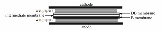 Figure 1. Experimental set-up for DB.
(a) Primary antibodies: monoclonal mouse anti-human EPO.
(b) Secondary antibodies: biotinylated goat anti-mouse IgG(H + L).
(c) Amplifying systems: Streptavidin:biotinylated peroxidase complexes.
(d) Development system: Chemiluminescence.
(e) Blocking buffer: 5 % (w/v) nonfat milk in PBS buffer.
Reference