
Inquiry Basket
 Loading ......
Chronic infection with HBV can lead to life-threatening liver diseases, such as cirrhosis and liver cancer. HBV is hepatotropic, and is a DNA virus. HBV is a known carcinogen. The nucleocapsid core of HBV is enclosed by a lipoprotein membrane. The virus encodes 3 major structural antigens: HBsAg, HBcAg, and HBeAg. HBV is classified, on the basis of antigenic epitopes of the envelope proteins, into 4 major serotypes. HBV has 10 major genotypes, which differ by more than 8% in their full genome nucleotide sequences. Genotypes have different virological features and geographic distribution. For instance, genotype A is the most prevalent genotype in North America, Northwestern Europe, India and Africa, while genotypes B and C are the most common genotypes in East Asian countries.
The HBV genome is made up of relaxed circular DNA (rcDNA) with an incomplete positive strand and a complete negative strand. It contains four open reading frames (ORFs), which overlap, named C, P, S, and X. The C ORF includes both the precore and core regions, and encodes HBc and HBe, depending on the start site of translation. The P ORF encodes a large polymerase protein (Pol) of around 800 amino acids through three functional domains; a terminal protein domain, a reverse transcriptase domain, and an RNase H domain. The S ORF is made up of the pre-S1, pre-S2 and S regions, encoding the three surface antigens L-HBs, M-HBs, and S-HBs. The X ORF encodes a 16.5 kDa protein, called HBx, with many functions in signal transduction, transcription activation, and DNA repair.
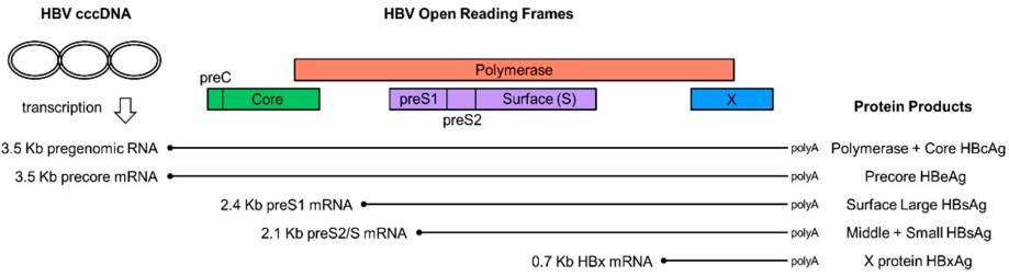 Figure 1. Hepatitis B virus gene expression
(Source: Hadziyannis E, et al. 2018)
Post HBV infection, rcDNA is transformed into covalently closed circular DNA (cccDNA), which persists as an epigenetically active viral genome. cccDNA is the major persistent form of the virus. The cccDNA complex is linked to host proteins to form a viral minichromosome, which is the template for all viral RNA transcription. The cccDNA generates HBV RNAs of different lengths.
The diagnosis and follow-up of HBV infection rely on laboratory viral biomarkers. HBV biomarkers are largely classified into two groups. The first group of biomarkers is the serological tests, which include detection and quantification of viral antigens and virus-specific antibodies. The second group is the nucleic acid tests, which are used for detection and quantification of HBV genome and its RNA transcripts. Both of these tests are commonly performed to diagnose acute and chronic HBV infection and to monitor chronic HBV infection. The serological tests for HBeAg and its corresponding antibody are the most common tests used for the diagnosis of HBeAg-negative CHB. The most widely used clinical serum biomarkers are quantitative HBsAg, HBeAg, HBV DNA, and ALT levels. HBsAg is the most clinically significant marker of HBV infection. Serum HBsAg detected for ≥6 months indicates chronic infection. HBsAg-positive individuals are regarded as infectious. HBeAg is positive during the early active replication phase of infection, indicating high infectivity. Some patients may remain HBeAg positive for many years. By detecting combinations of serological markers such as HBsAg, HBeAg, their antibodies, and anti-core antibodies, it is possible to determine whether a patient is in an acute, chronic, immune, or susceptible state.
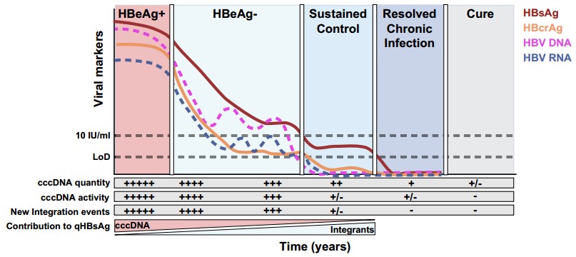 Figure 2. Proposed new definitions of the stages of HBV control
(Source: Feld JJ, et al. 2025)
HBsAg was the first discovered biomarker of HBV infection. It remains the most commonly used diagnostic test for HBV infection to this day. HBsAg is translated from mRNA transcribed from cccDNA and/or HBV sequences that have become integrated into the host's genome. HBsAg is overproduced in HBV infected liver cells, and secreted into the serum in great quantities. It is thus a highly sensitive and specific biomarker for HBV infection. In the majority of cases, it is enough to diagnose acute HBV infection and screen for chronic HBV infection based on the results of the HBsAg test alone, together with a concomitant anti-HBs antibody test and anti-HBc antibody test.
Table 1. Hepatitis B Virus (HBV) serology interpretation
| HBsAg | Total Anti-HBc | IgM Anti-HBc | Anti-HBs | Interpretation |
| - | - | - | - | No HBV infection - susceptible |
| - | - | - | + | HBV immune - vaccinated |
| - | + | - | + | Past HBV infection |
| + | + | + | - | Acute HBV infection |
| + | + | - | - | Chronic HBV infection |
| - | + | - | - | Inconclusive |
(Source: Hadziyannis E, et al. 2018)
Assays for HBeAg and anti-HBe antibody are done after chronic HBV infection has been diagnosed, and these are often measured by the same assays used for the previously described serologic markers. In the HBeAg-positive phase of infection, levels of HBsAg are strongly correlated with serum HBV DNA and with intrahepatic cccDNA, while the correlation is poor in the HBeAg-negative phase. This is partly because the source of HBsAg shifts - from mainly deriving from cccDNA to more from integrated HBV DNA. Additionally, HBeAg-positive patients have higher HBsAg titers than HBeAg-negative patients.
HBeAg is a non-structural protein translated from the precore mRNA, processed in the endoplasmic reticulum, secreted extracellularly, and circulates in the blood. Although HBeAg is not essential for viral replication or infection, it plays a key role in virus-host interactions and the establishment of chronic HBV infection. This non-structural protein is highly conserved among all orthohepadnaviruses and has mainly been regarded as an auxiliary protein of HBV, used clinically as a marker of active viral replication. HBeAg correlates significantly with intrahepatic HBcAg detection, but a notable proportion of HBeAg-negative/anti-HBe-positive carriers show evidence of unexplained chronic hepatitis without intrahepatic HBcAg. Only some of these patients have co-infection with HDV, which does not explain all cases. Further molecular diagnostic analysis revealed the presence of HBV DNA in the serum of all HbeAg negative and positive patients with intrahepatic HBcAg positivity. Serum HBV DNA levels are lower in HBeAg-negative patients than in HBeAg-positive patients. HBeAg seroconversion (loss of HBeAg and appearance of anti-HBe) occurs at an annual rate of approximately 10%–20% of patients. HBeAg positivity is usually a marker of active viral replication and high infectivity; HBeAg negativity is usually a marker of reduced viral replication, although regular monitoring is necessary as reactivation or progression to HBeAg-negative hepatitis can occur. Quantitative HBeAg (qHBeAg), which can be measured by laboratory-developed methods during HBeAg-positive infection, is a potential biomarker. Monitoring qHBeAg helps predict treatment response and sustained HBeAg seroconversion. Studies found that patients with qHBeAg levels remaining >10 kU/mL from baseline to the end of treatment had a 100% negative predictive value for HBeAg seroconversion.
HBeAg has multiple immunoregulatory functions, including downregulating TLR2, NF-κB activation, and IL-18-mediated IFNγ expression signals, which promote viral replication. HBeAg weakens the adaptive immune response by preferentially activating Th2 cells rather than Th1 cells. Th2 cells induce non-protective humoral responses, while Th1 cells stimulate macrophages to eliminate viral particles. HBeAg in serum shifts the normal Th1-mediated anti-HBc antibody response toward a Th2 phenotype. Several studies have identified that HBeAg can also modulate macrophage metabolism and function. It was reported that HBeAg could augment OXPHOS in macrophages in a TLR4-dependent manner, and it could also induce the expression of death receptor 5 (DR5) and death-associated protein 3 (DAP3) to mediate the aforementioned effect. HBeAg could also induce pyroptosis and apoptosis in M1-like macrophages and could induce mainly apoptosis in M2-like macrophages. The findings from the study could offer a mechanistic basis for metabolic reprogramming of macrophages by HBV to attenuate their antiviral response.
References
| Target | Cat. No. | Product Name | Size | Species | Application | Detection Sample | |
| HBsAg | DEIA001 | Hepatitis B Surface Antigen ELISA Kit | 96T | Human | Qualitative | Blood | Inquiry |
| IVDEIA001 | Human Hepatitis B surface antigen (HBsAg) ELISA Kit | 96T | Human | Qualitative | Serum, plasma | Inquiry | |
| DEIA-JY2436 | HBsAg Glycan ELISA Kit | 96T | Human | Quantitative | Cell culture supernatant, serum, and plasma | Inquiry | |
| DEIASL263-2 | Mouse Anti-HBsAg IgG ELISA Kit | 96T | Mouse | Quantitative | Serum | Inquiry | |
| DEIA-NS2502-4 | Hepatitis B Surface Antigen (HBsAg) ELISA Kit | 96T | HBV | Quantitative and qualitative | Cell culture, bioprocessing solutions, and or other samples (e.g., blood, saliva, mucosa) | Inquiry | |
| HBeAg | DEIA003 | Human HBeAg ELISA Kit | 96T | Human | Qualitative | Serum, plasma | Inquiry |
| DEIA004 | Human Anti-Hepatitis B Virus E Antigen (HBe) Antibody ELISA Kit | 96T | Human | Qualitative | Serum, plasma | Inquiry |
Loading ......
