
Inquiry Basket
 Loading ......
Anti-programmed cell death 1 (PD-1) antibodies are in wide use for the treatment of various cancers, particularly cancers with a high tumor mutation burden (TMB) such as advanced cutaneous melanoma. Lpilimumab is a fully humanized immunoglobulin (Ig)G1 monoclonal antibody that blocks cytotoxic T-lymphocyte antigen (CTLA-4) to activate and increase T cells, particularly the tumor recognized T-cell clones that reside in primary tumors. Combination therapy comprising nivolumab and ipilimumab or sequential administration of nivolumab and ipilimumab with a planned switch are among the most effective chemotherapies against advanced melanoma, and even increase the response rate (RR) for untreated metastasis of melanoma to the brain compared to nivolumab monotherapy. This review focuses on the recent development of biomarkers for assessing the efficacy of anti-PD1 antibodies using routine blood tests such as the neutrophil-to-lymphocyte ratio, eosinophil ratio, serum markers such as lactate dehydrogenase (LDH), PD-L1 expression on melanoma cells, microsatellite instability (MSI) and mismatch repair deficiency assays, as well as soluble CD163, and tumor-associated macrophage (TAM)-related chemokines (e.g., CXCL5, CXCL10).
More about chemokines and chemokines receptors
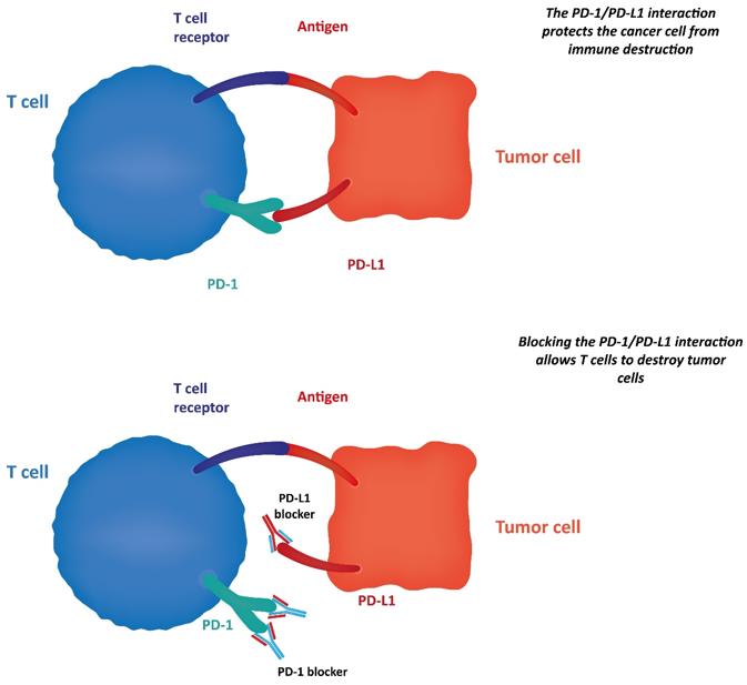 Fig 1. The PD-1/ PD-L1 Blockade
Recent reports have suggested the significance of routine blood tests, such as cell counts and cell ratios, for predicting the efficacy of anti-PD1 antibodies against advanced melanomas. Baseline NLR could be one possible predictive marker for immune checkpoint inhibitor (ICI)-treated patients with advanced melanoma. Lower ALC shows significantly less clinical benefit from anti PD1 antibody, which is associated with pretreatment NLR in patients with head and neck squamous cell carcinoma. Overall, these reports have suggested that baseline routine blood tests are important for predicting the efficacy of ICI. Generally, large baseline tumor size in parallel with increased levels of LDH correlates with poor prognosis in advanced melanoma patients. Indeed, patients with elevated baseline LDH showed significantly shorter OS compared to patients with normal LDH. ECOG performance status (PS) and elevated LDH were reported as independent variables significantly associated with poor OS. Increased LDH correlated with the poor prognostic factors of not only cutaneous melanoma, but also uveal melanoma, which possesses a high potential for rapid metastasis, and NSCLC. Since anti-PD1 antibody applies to various cancers, including gastric cancer, renal cell carcinoma, and Hodgkin lymphoma, measurement of LDH might offer a useful, standard marker for patients treated using ICI.
In cutaneous melanoma, both tumor cells and TAMs express PD-L1, leading to the maintenance of an immunosuppressive microenvironment at tumor sites. Accordingly, many studies have suggested that PD-L1 expression on melanoma cells can represent a biomarker for predicting the efficacy of anti-PD1 antibodies, and even other ICIs. Although those reports suggested the clinical benefits of assessing PD-L1 expression on melanoma cells in predicting the clinical outcomes of ICI treatment, the clinical utility in the real world is limited because of the low sensitivity of immunohistochemical (IHC) assays using different antibody clones, staining platforms and scoring systems in each institute
The high RR to anti-PD1 antibodies for cancers with high frequency of MSI has been highlighted in many recent clinical studies. Among cancer species, colorectal cancer and endometrial cancer possess a high frequency of MSI, leading to the results of clinical studies that have presented significantly improved RR, PFS and OS in patients with mismatch-repair deficient colorectal cancers compared to those of mismatch repair-proficient colorectal cancers. Since high expression of PD-L1 can provide a biomarker for predicting the efficacy of anti-PD1 antibodies, a high frequency of MSI could correlate with RR, PFS and OS following use of anti-PD1 antibodies. High TMB correlated with increased neoantigens in various cancers, and could provide predictors for the efficacy of ICI treatment.
TAMs are functionally reprogrammed to polarized phenotypes by exposure to various factors, leading to the maintenance of a tumor microenvironment. Expression of PD-L1 on TAMs is modified by both stromal factors such as regulatory T cells (Tregs) and exogenous factors including immune therapies. Since PDL1 on tumor cells could be one prognostic factor for melanoma patients treated with ICIs, TAM related factors could offer biomarkers for predicting the efficacy of ICI. TAMs in melanoma patients express not only PD-L1, but also PD-1. Because PD-1 expression in TAMs is one of the key factors in M2 macrophage polarization, administration of an anti-PD1 antibody might repolarize TAMs, leading to TAM activation in melanoma patients. Those reports suggested that serum sCD163 could offer a predictive marker for the efficacy and irAEs of antiPD1 antibodies. In addition to sCD163, TAM-related chemokines could provide another group of prognostic markers for the outcomes of anti-PD1 antibody treatment. Although combination therapy with nivolumab and ipilimumab is recommended by the NCCN guideline for cutaneous melanoma as a first-line therapy, this combination therapy leads to a high frequency of SAEs among patients with advanced melanoma. In addition to sCD163, TAM-related chemokines could provide another group of prognostic markers for the outcomes of anti-PD1 antibody treatment (50). For example, CXCL5 is a chemokine that can recruit neutrophils
References
| Target | Cat. No. | Product Name | Species Reactivity | Size | Application | Detection Sample | |
| PDCD1 | ABPR-0664 | Human PDCD1 ELISA Matched Antibody Pair | ELISA | Inquiry | |||
| DEIA-BJ357 | Human Soluble programmed death-1 ELISA kit | Human | 96T | Quantitative | Serum, plasma, tissue homogenates and other biological fluids | Inquiry |
Loading ......
