
Inquiry Basket
 Loading ......
Immunoglobulin E (IgE) was discovered in the mid-1960s and has been proved to play an important role in type I allergic reactions. The conversion of antibody homotype to IgE may occur in nasal mucosa, bronchial tissue, and intestinal mucosa. Dendritic cells (DC) are present in the epithelium and upper lamina propria of respiratory tract, intestinal tract, and skin, which can capture allergens and promote T cells to polarize into allergic type II immune response. B cells are also important antigen presenting cells (APC) that initiate IgE hypersensitivity. The increased expression of Th2 cytokines such as IL-4 and IL-13 produced by mast cells and basophils in nasal mucosa can indirectly promote tissue mast cells to induce B cells to synthesize IgE, which leads to local synthesis of IgE by B cells. There is evidence that allergen-specific antibodies (non-IgE) in early life can regulate allergic sensitization. Maternal immunoglobulin is transferred to offspring during pregnancy and through breast milk, and maternal-specific allergen IgG seems to protect offspring from allergies. The recovery of immune tolerance after long-term allergen immunotherapy is related to the induction of local and systemic neutralizing antibodies against IgG and IgA.
IgE is structurally identical to other antibody classes, consisting of two identical light and heavy chains. Each chain consists of 110 amino acids that make up the "immunoglobulin structural domain", with disulfide bonds covalently connecting the light and heavy chains. Different from IgD, IgG and IgA with three constant region domains, the heavy chain is similar to the μ heavy chain of IgM in structure because it has four constant region domains (Cε1-Cε4). Cε3 and Cε4 domains are homologous to Cγ2 and Cγ3 domains of IgG antibody in sequence and quaternary structure. The difference between IgE and IgG lies in the position of its Cε2 domain, which is inserted into the corresponding Fab-Fc hinge region of IgG. The two antigen binding sites are formed by pairing the variable regions of the light chain and the heavy chain. IgE bends asymmetrically at the Cε2-3 linker, folds in on itself, and the two Cε2 structural domains fold backward, almost touching the Cε4 structural domain.
Immunoglobulin E is the core of type I immediate anaphylaxis. Several studies have shown that allotype conversion of antibodies beneficial to IgE occurs in the nasal and bronchial mucosa of allergic patients and in lymphoid tissue near the contact site of allergens. The concentrations of IgE antibody and FcεRI in target organs were increased, and the concentrations of atopic and allergic patients were more than ten times higher than those of non-atopic patients. IgE antibody has high affinity with FcεRI on the surface of mast cells and basophils.
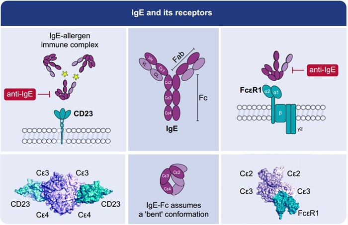 Figure 1. IgE and its receptors
(Source: Shamji MH, et al. 2021)
The high-affinity IgE receptor (FcεRI) is a member of the immunoglobulin (Ig) superfamily. It is highly expressed on the surface of mast cells and basophils as an αβγ2 tetramer (~200,000 molecules/cell). It consists of four polypeptide chains, an α chain, a β chain and two disulfide-linked γ chains. In human monocytes, Langerhans cells, peripheral blood dendritic cells, eosinophils, platelets, and smooth muscle cells, FcεRI is expressed in the form of αγ2 trimer, which consists of one α and two γ chains. The alpha chain consists of an extracellular domain, a transmembrane helical domain, and a short cytoplasmic sequence. The IgE binding function of high affinity IgE receptor is limited to the two extracellular domains of the α chain, with a binding ratio of 1:1.
The intracellular sequences of both the β and γ chains consist of immunoreceptor tyrosine-based activation motifs (ITAMs), and the β subunit chain amplifies downstream events upon initial activation of the surface FcεRI. Trimeric expression of FcεRI on monocytes, DCs, and Langerhans cells has been shown to promote allergen presentation to CD4+ T cells. FcεRI-mediated uptake of allergens by APCs is 100 to 1,000 times more efficient than any endocytosis or phagocytosis.
In the presence of IgE, both types of cells are activated by high affinity IgE receptors. Allergen-induced IgE cross-linking with FcεRIs on the surface of mast cells or basophils induces receptor aggregation and intracellular signal events, which leads to Ca2+-dependent release of formed mediators and the re-synthesis and secretion of lipid mediators and cytokines such as IL-4 and IL13. The concentration of allergen-specific IgE, IgE affinity and the ratio of allergen specificity to total IgE are the key factors to determine the release intensity of effector cells.
 Figure 2. IgE-mediated Th2 and Mast cell/basophil activation and inhibitory effects of allergen-specific IgG and IgA as well as anti-IgE.
(Source: Shamji MH, et al. 2021)
Studies have shown that the specific IgE-allergen complex can significantly enhance the allergen-specific T cell response at low allergen concentrations. This IgE-mediated allergen presentation or promotion of allergen presentation involves the binding of IgE-allergen complex to CD23 on antigen-presenting B cells. It was also found that dendritic cells and monocytes in peripheral blood expressed high affinity IgE receptor (FcεRI) and activated allergen-specific T cells in an IgE-induced manner. In fact, these studies are related because the level of allergen in the respiratory tract is extremely low during natural exposure to allergens, and in this case of hypoallergen exposure, the presentation of allergens to T cells promoted by IgE activates T cells.
The binding of allergen-IgE complex to antigen-presenting cells depends on many parameters, such as the antigen specificity and affinity of IgE antibody, the expression level of receptor and the clonality of B cells. In addition, the complexity and affinity of IgE binding to multiple epitopes on allergens have been shown to be related to the promotion of T cell response. These results suggest that the number of IgE molecules bound to each allergen may play an important role in the formation and binding of this complex.
Although IgE-mediated type I hypersensitivity usually leads to persistent inflammation, on the other hand, IgE-mediated type I hypersensitivity is obviously beneficial to the host in terms of inflammatory process, especially in defense against various microbial pathogens. For example, mast cells play a protective role in soil-borne Helminthes, while basophils play a central role in preventing tick bites. From snakes to reptiles to arthropods, the importance of immunoglobulin-mediated type I hypersensitivity in defending against the venom of a variety of organisms has also been confirmed. These protective effects involve mast cell protein hydrolases such as chymase, tryptase, and carboxypeptidase A3, which are stored in granules and can rapidly degrade and inactivate toxic peptides. A series of studies have shown that allergic reactions mediated by IgE also contribute to acquired resistance to potentially fatal effects of venom, such as allergic reactions induced by honeybee venom. Although this reaction can be fatal in some "unfortunate" individuals, it also helps protect the host by inactivating the venom by releasing proteases.
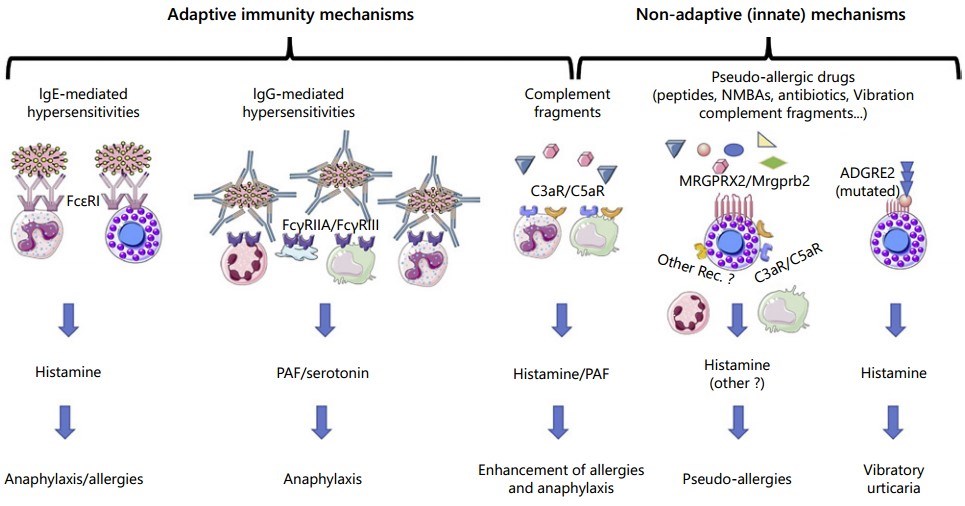 Figure 3. Summary of described hypersensitivity mechanisms
(Source: Vitte J, et al. 2022)
References
| Target | Cat. No. | Product Name | Size | Species | Application | Detection Sample | |
| IgE | DEIAGEF-01 | Food Allergen specific IgE ELISA Kit | 8T | Human | Qualitative | Serum | Inquiry |
| DEIAGEI-01 | Inhalant Allergen specific IgE ELISA Kit | 8T | Human | Qualitative | Serum | Inquiry | |
| DEIA632 | Dog IgE ELISA Kit | 96T | Dog | Quantitative | Serum, plasma | Inquiry | |
| DEIA-BY041 | Monkey IgE ELISA Kit | 96T | Monkey | Quantitative | Serum | Inquiry | |
| DEIA9467 | Human IgE ELISA Kit | 96T | Human | Quantitative | Serum | Inquiry | |
| DEIA06101 | Rat IgE ELISA kit | 96T | Rat | Quantitative | Serum, plasma, culture medium | Inquiry | |
| DEIA652 | Mouse IgE ELISA Kit | 96T | Mouse | Quantitative | Serum, plasma | Inquiry | |
| DEIA268 | Total IgE ELISA Kit | 96T | Quantitative | Serum, plasma | Inquiry | ||
| DEIABL50 | Total human IgE ELISA Kit | 96T | Quantitative | Plasma, Serum | Inquiry | ||
| DEIA652V2 | Mouse IgE ELISA Kit | 96T | Quantitative | Biological samples | Inquiry | ||
| DEIACL1 | CDSimple™ IgE Chemiluminescent ELISA Kit | 96T | Quantitative | Serum | Inquiry |
| Target | Cat. No. | Product Name | Host | Isotype | Application | |
| IgE | DAGIC1432 | Mouse IgE Isotype Control | Mouse | IgE | FC, EIA, WB | Inquiry |
| DAG-IC98 | Mouse IgE | Mouse | IgE | ELISA, FC, WB | Inquiry | |
| CABT-L2425 | Anti-Human IgE chimeric monoclonal antibody, clone 25I2 | Mouse | IgA | ELISA | Inquiry | |
| DCAB-TJ280 | Magic™ Anti-Human IgE monoclonal antibody, clone 2B3 [HRP] | Mouse | IgG1 | EIA, ELISA(Det) | Inquiry | |
| DCABH-8940 | Anti-Human IgHE monoclonal antibody, clone CF6 [FITC] | Mouse | IgG1 | FC | Inquiry | |
| CABT-L2426 | Anti-Human IgE chimeric monoclonal antibody, clone 25I9 | Mouse | IgG | ELISA | Inquiry | |
| DMABP-L13 | Magic™ Anti-Human Immunoglobulin E (IgE) Monoclonal Antibody, clone GB428 | Mouse | IgG2b | ELISA | Inquiry | |
| DMABP-L14 | Magic™ Anti-Human Immunoglobulin E (IgE) Monoclonal Antibody, clone GB429 | Mouse | IgG2b | ELISA | Inquiry | |
| DMABP-L15 | Magic™ Anti-Human Immunoglobulin E Monoclonal Antibody, clone IN228 [HRP] | Mouse | IgG | ELISA | Inquiry | |
| DCABH-84 | Anti-Human IgHE monoclonal antibody, clone 5D4 | Mouse | IgG1 | WB, ELISA, sELISA | Inquiry | |
| IgE kappa | DAGIC800 | Mouse IgE kappa Isotype Control | Mouse | IgE, κ | EIA, FC, WB | Inquiry |
| Target | Cat. No. | Product Name | Expression System | Tag/Conjugate | Application | |
| IgE | DAGF-100 | Purified Dog IgE positive control | Dog | Unconjugated | Inquiry | |
| DAGA-828 | Immunoglobulin E (>98%) | Human myeloma plasma | Unconjugated | Controls, Calibrators, ELISA, Blotting | Inquiry |
Loading ......
