
 Reagents for Rabies Virus Research
Inquiry Basket
 Loading ......
Lyssavirus, a genus of the family Rhabdoviridae from order Mononegavirales, consists of single-stranded, negative-sense RNA viruses, which infect mammals and cause fatal acute viral encephalomyelitis known as rabies[1]. Rabies virus the prototype virus of the genus Lyssavirus, is more globally distributed and abundant with more known progenies amongst species in the genus[1]. Unfortunately, there is no effective therapy for rabies once the symptoms of clinical disease occur. Annually, about 60,000 human rabies deaths are reported worldwide. However, the disease is 100% vaccine-preventable through the prompt administration of human postexposure prophylaxis (PEP) and vaccination of animal reservoirs. The first rabies vaccine was introduced in 1885 and was followed by an improved version in 1908. It is on the World Health Organization's List of Essential Medicines.
RABV is a negative-stranded RNA virus of the Rhabdoviridae family. RABV virions [G] are enveloped by a host cell-derived membrane and take on a bullet shape of about 200 nm by 80 nm. The bullet shape is likely influenced by the constraints of budding and viral uptake.
The relatively small RNA genome of the virus (~12 kb) encodes for five proteins: nucleoprotein (N), phosphoprotein (P), matrix protein (M), glycoprotein (G), and polymerase (L). RABV virion has two main structural components including an envelope and a helical ribonucleoprotein core (RNP). N proteins encapsidate the genomic and antigenomic RNA. The resulting ribonucleoprotein (RNP) complex protects the RNA from degradation by RNAses. The phosphoprotein provides the connection between the RNP and viral polymerase protein, and antagonizes innate immunity in the infected host. The matrix protein coats the inside of the viral envelope, bridging the carboxy-terminal region of the glycoprotein and the RNP. Importantly, it sequesters the glycoprotein in a concentrated area of the cell membrane to aid in viral budding. The glycoprotein interacts with a host cell receptor and mediates pH-triggered fusion between the viral and host membranes, which results in the release of the RNP into the host cytoplasm. The glycoprotein is also the primary target for virus-neutralizing antibodies, which are necessary for protection against rabies disease. The virus requires its own polymerase to perform the primary transcription of mRNA from the anti-genomic RNP and initiate the viral life cycle within the infected cell. After a threshold of transcription has been achieved and a certain amount of viral proteins has been produced, the polymerase switches to replication mode in which it ignores stop and start signals used to produce single mRNAs and produces a single, positive-stranded encapsidated plus-sense RNA. This anti-genomic RNA serves as a template for more copies of the genomic viral RNA. Such newly produced RNP are assembled together with the matrix protein and the glycoprotein into new virions at the cell membrane [1, 2].
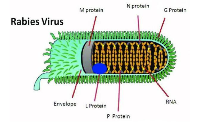 Fig 1. Structure of Rabies virus
In the first phase of the rabies virus (RABV) life cycle, the virus binds to the cell surface receptors via its glycoprotein and enters by endocytosis. Subsequently, the viral membrane fuses with the endosomal membrane to release the viral genome. In the second phase, the encapsidated negative-stranded RNA genome is transcribed by the polymerase complex, starting with a short uncapped leader RNA (leRNA), followed by the transcription of 5′ end-capped (cap) and polyadenylated (A) mRNAs, and their translation into the viral proteins nucleoprotein (N), phosphoprotein (P), matrix protein (M), glycoprotein (G) and polymerase (L). Following replication, the full-length antigenomic RNA is encapsidated in the nucleoprotein protein along with the genomic RNA. The synthesized antigenome functions as a template for the synthesis of additional copies of genomic RNA. In the last phase, the viral components are assembled and the RABV virions bud and are released, starting a new round of infection [3].
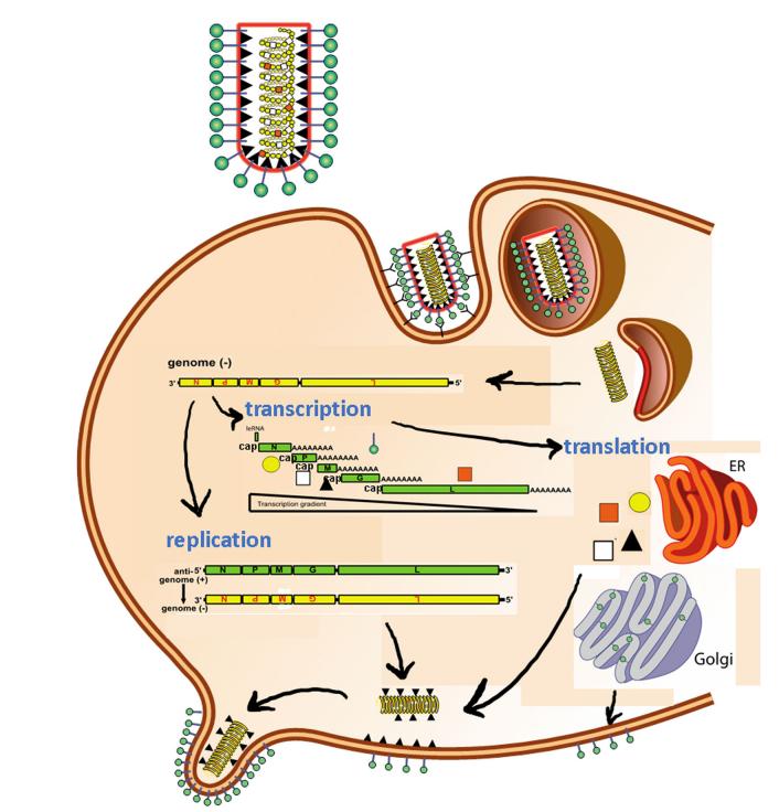 Fig.2 Life cycle of Rabies virus
The rabies life cycle and its resulting pathogenesis have been extensively studied and reviewed. Briefly, RABV, with its modest genome and single surface glycoprotein, can infiltrate an astonishing number of mostly neuronal tissues in almost any mammal to induce its lethal pathology. It has been suggested that the nAchR receptor enriches RABV at the neuromuscular junction (NMJ, synaptic cleft), enabling more efficient infection of the connected motor neurons. Initial virus amplification occurs in muscle, which indicates that nAchRs might be used to infect muscle cells. RABV can enter neurons by binding to NCAM or another, unknown receptor. Following uptake by clathrin-mediated endocytosis, RABV virions are then transported within the vesicle and are released in the cell body of the infected neurons, where replication and transcription occurs [4, 5].
Infection usually begins in muscle tissue following a bite from an infected animal. The virus then crosses neuromuscular junctions to peripheral nerves and uses retrograde axonal transport to reach the central nervous system (CNS). Replication occurs in some instances in muscle. However, strong immune evasion may make it difficult to detect rabies virus in early infection [6].
Whereas the initial spread of RABV is exclusively retrograde, at the end stage of infection, RABV reverses its direction of transport and migrates out of the CNS in an anterograde fashion towards the periphery. Centrifugal spread, especially to the salivary glands where the virus can easily transmit through a bite, is essential to restart the cycle. Rabies disease then rapidly deteriorates the health of the host and leads to death from respiratory or heart failure, usually within days [7].
Rabies vaccination was invented in the late 19th century and improved over the 20th century. Upon first review, there appears little incentive for further improvement: current vaccines work extremely well, providing safe, longlasting protection against the lethal disease.
The first vaccine against RABV was created in 1885 by Louis Pasteur who, despite not yet knowing about the infectious agent itself, accurately traced the infection to the brain and spinal cord in rabies-infected rabbits. By the 1950s and 1960s, non-neuronally derived vaccines are produced. First, purified duck embryo vaccines (PDEVs) became available. Then, in vitro cell culture was developed and perfected, enabling the most important innovation in RABV vaccines: growth on human diploid cells. There are four general varieties based on production method: the human diploid cell vaccine (HDCV), purified chick embryo cell vaccine (PCECV), primary hamster kidney cell vaccine (PHKCV) and, most recently, purified Vero cell rabies vaccine (PVRV). Since the 1980s, the development of vaccines for domestic and wild animals has progressed even further, utilising alternative viral vectors and oral administration [8].
Despite the successful track record, rabies vaccines need to be improved. They have two primary limitations: they require multiple doses to achieve high neutralising titres in all recipients, and they are costly. Another limitation of the vaccine is the lack of coverage against nonRABV lyssaviruses. Given that rabies continues to be a problem in large parts of the world despite the existence of functional rabies vaccines, new innovations are the only way to reduce rabies disease to acceptable rates [8].
The ideal rabies vaccine would be a safe, inexpensive, single-dose vaccine that is temperature-stable for long periods of time and is easy to administer. For a realistic picture of how close we are to approaching this ideal, one can look to the pipeline of clinical trials for rabies therapeutics to see what improvements may be approved in the near future. The expansion of the vaccine market to new countries or populations, the introduction of a new administration schedule, the administration of the rabies vaccines with other vaccines, or the administration of the rabies vaccine to immunocompromised individuals, such as, A serum-free PVRV containing no components of human or animal origin and without antibiotics. The vaccine, which has completed phase III trials, reduces the risk of contamination by serum-transmissible elements.
References
| Cat. | Product Name | Expression System | Application | |
| DAGC695 | Native RABV Antigen | Human diploid cells | Immunogen, WB, ELISA | Inquiry |
| DAGF-021 | Recombinant RABV Glycoprotein (aa 20-458)[His] | E. coli | WB, Standard, ELISA, Immunogen | Inquiry |
| DAG2309 | Recombinant RABV M2 Protein (a.a. 1-202) [His] | E. coli | WB, ELISA | Inquiry |
| DAG2364 | Recombinant RABV Nucleoprotein (a.a. 1-450) [His] | E. coli | ELISA, WB, Immunogen | Inquiry |
| Cat. | Product Name | Species Reactivity | Detection Sample | |
| DEIAHRVPY28 | Human Anti-RABV Glycoprotein IgG ELISA Kit | Human | Serum, Plasma or other biological fluids | Inquiry |
| DEIA-RV2310-6 | Rabbit Anti-RABV IgM ELISA kit | Rabbit | Serum | Inquiry |
| DEIA-RV2310-7 | Monkey Anti-RABV IgG ELISA Kit | Monkey | Serum, plasma or other biological fluids | Inquiry |
| DEIA-RV2310-9 | Rabbit Anti-RABV IgG ELISA Kit | Rabbit | Serum | Inquiry |
| DEIA-RV2310-14 | G. Pig Anti-RABV IgG ELISA Kit | G. Pig | serum, plasma | Inquiry |
| DEIA-RV2310-15 | RABV Vaccine ELISA Kit | N/A | Vaccines formulated in Aluminum hydroxide or Alum gel | Inquiry |
| DEIA-RV2310-17 | Human Anti-RABV NP IgG ELISA Kit | Human | Serum, Plasma or Biological Samples | Inquiry |
| DEIA-RV2310-18 | Mouse Anti-RABV NP IgG ELISA Kit | Mouse | Serum, Plasma or Biological Samples | Inquiry |
| DEIA-RV2310-19 | Rabbit Anti-RABV NP IgG ELISA Kit | Rabbit | Serum, Plasma or Biological Samples | Inquiry |
Loading ......
