
Inquiry Basket
 Loading ......
Varicella-zoster virus (VZV) virus is a virus containing double-stranded DNA. Its morphological structure is polygonal or round, and its entire diameter is 80-120nm. VZV has a lipid-rich envelope, which is obtained from the cell membrane, and into which the virus's own glycoproteins are inserted. The lower layer of the envelope is the sheath, which contains proteins and enzymes. It is surrounded by an icosahedral symmetrical nucleocapsid, which contains the core of the linear double-stranded DNA genome in the middle. VZV is the smallest herpesvirus, with a genome of approximately 125,000 bp containing two special segments that are covalently linked and surrounded by inverted repeat regions. 71 open reading frames (ORFs) were initially predicted in its genome, and transcripts of all these protein-coding regions were detected during viral infection, including 8 glycoproteins, namely gE, gB, gH, and gI, gK, gN, gC and gL. The VZV genome is genetically quite stable, with a relatively low nucleotide mutation rate. Based on the single nucleotide polymorphism (SNP) spectrum of multiple open reading frames (ORFs) of the virus, 5 major VZV genetic clades (clades 1 to 5) and 2 tentative genetic clades (VI and VII).
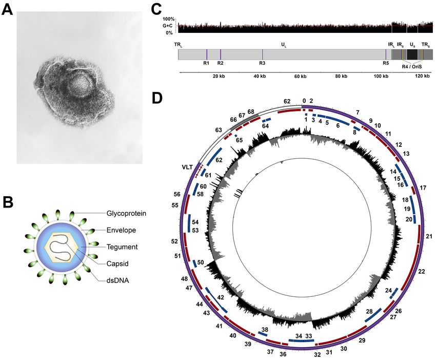 Figure 1. Structure of varicella-zoster virus (VZV) particles and genome. (Depledge DP, et al.; 2018)
VZV replicates in a variety of human cells but remains latent in neurons. The virus acquires neurons by hematogenous dissemination or retrograde axonal transport from cutaneous varicella vesicles to ganglia. Unlike the latent period of herpes simplex virus, which is limited to cranial ganglia (HSV-1) or sacral ganglia (HSV-2), VZV latent in cranial ganglia, dorsal root ganglia, and autonomic ganglia along the entire neural axis. Once inside the neuron, the viral DNA enters the nucleus, circulates, and binds to histones in the ice-bathed cell, after which viral gene transcription is restricted.
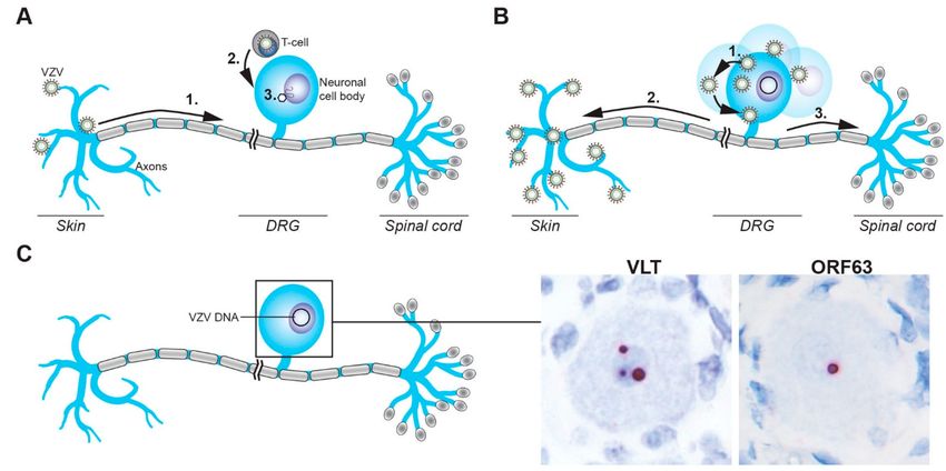 Figure 2. Schematic representation of the establishment of, and reactivation from, VZV latency in sensory neurons. (Depledge DP, et al.; 2018)
Because VZV is latent during primary varicella, similar to the Oka strain of attenuated VZV used for varicella vaccination, almost everyone has latent VZV. The latent period is characterized by the presence of viral DNA in the body, but limited expression of viral genes, thus preventing the production of infectious viruses. The latent phase of the VZV virus is characterized by the fact that it can be reactivated and, in most cases, produce more severe disease than the initial infection.
There are 9 glycoproteins on the VZV viral envelope, namely gE, gI, gC, gH, gL, gB, gK, gM and gN. gE is encoded by ORF68 and is the most abundant protein expressed on the surface of infected cells. It is a transmembrane protein containing 623 amino acids and is divided into four parts: signal peptide, extracellular region, transmembrane region and intracellular region. It is a key protein for VZV replication and virus assembly, and is responsible for mediating the spread of the virus between cells. At the same time, gE is the main target of virus-specific antibodies and T cell responses, is highly immunogenic, and is often used as a candidate antigen for vaccine production. gI is encoded by ORF67 and is often linked to gE to form a heterodimeric gE/gI complex. This complex can act as an Fc receptor on infected cells. gC is encoded by ORF14, and only a small amount of gC is synthesized during the first 3 to 4 cycles of VZV replication. Depending on the titer of the infecting virus, the presence of gC can be detected 48 to 72 hours after vaccination, and its expression level can reflect the virulence of VZV. gH is encoded by ORF37 and can be used as an important antigen in vaccine preparation. It contains a potent neutralizing epitope and induces the production of neutralizing antibodies. gB is encoded by ORF31 and can elicit neutralizing antibody responses similar to but weaker than gH. gL is encoded by ORF60, which can turn gH into a glycosylation product, which is a prerequisite for the maturation of gH. Mature gL plays a role similar to a molecular chaperone in the gH/gL complex. gK, encoded by ORF5, is a hydrophobic protein with multiple membrane-binding domains and is relatively conserved among alphaherpesviruses. gM is encoded by ORF50, and gN is encoded by ORF8.5 or ORF9A. They are newly discovered proteins in recent years. gN is present in the cell membrane of infected cells.
PCR is one of the main methods for detecting viral nucleotides. Viral RNA can also be identified by reverse transcribing RNA into DNA, that is, reverse transcription PCR (RT-PCR). The real-time quantitative PCR method (qPCR) refers to a method that adds fluorescent groups to the PCR reaction system, uses fluorescence signal accumulation to monitor the entire PCR process in real time, and finally performs quantitative analysis of the unknown template through the standard curve. The fluorescent substances used in this method can be divided into two types: SYBRGreenⅠand TaqMan probes. The concentration of the virus in the detection sample is directly related to the initial copy number. When the fluorescence amplification signal reaches the threshold intensity, the Ct value is negatively related to the initial concentration of the virus in the sample. In actual detection, a standard curve can be made from a standard with a known starting copy number, and the Ct value of the unknown sample can be obtained through detection, and the starting copy number of the sample can be calculated.
The PCR method is used to detect the VZV viral load in patients' early blister fluid or peripheral blood, and is used as a laboratory method for the diagnosis of herpes zoster, which has been widely reported in clinical practice. When the quantitative PCR method is used to directly detect the virus titer in a sample, the infectivity of the virus cannot be distinguished from the DNA level, nor can the number of VZV viruses with infectivity be counted. Therefore, it is necessary to infect the cells with the sample and culture it for a certain period of time, so that the virus with the ability to infect is amplified in the cell. By detecting the content of the amplified viral DNA, the number of viruses with the ability to infect the original sample is estimated, and then the number of viruses with the ability to infect the sample is obtained.
This method can evaluate the virus titer from the DNA level, but its expensive equipment, high work intensity, high operator counting requirements and easy contamination limit its wide application. The timing of DNA extraction must be determined to ensure that the virus has infected the cells but the lack of large-scale replication is its technical barrier.
Western-Blot technology is based on the principle of specific binding of antigens and antibodies to detect certain proteins in complex samples such as cells, tissues, organs or body fluids. This method is based on SDS-PAGE, which separates various proteins according to their molecular weight, and then transfers the proteins to a nitrocellulose membrane through membrane transfer, binds and incubates them with specific antibodies, and finally uses certain indicator molecules (Such as horseradish peroxidase, alkaline phosphatase, etc.) to develop and analyze the target protein to achieve the purpose of qualitative and semi-quantitative analysis. Since this method is a qualitative and semi-quantitative analysis method with complex operation and high cost, it is mainly used in the identification and analysis of virus particle proteins in order to better understand the process and pathogenesis of virus infection in the body.
First developed as an improved method of traditional affinity column chromatography, it is a method of small-scale affinity purification of antigens using specific antibodies immobilized on solid supports such as magnetic beads or agarose resin. This method is one of the common methods for isolating proteins and other biomolecules from cell or tissue lysates for Western blot detection or other detection techniques.
The goal of immunoprecipitation is only to separate proteins sufficient for Western blot analysis or other semi-quantitative or quantitative detection methods. It is often used in conjunction with SDS-PAGE to determine the molecular weight and content of proteins.
The hemagglutination test is a method that uses the property of certain viral proteins (such as the agglutinin of influenza virus) to agglutinate with red blood cells (RBCs) to indirectly quantify viral particles. The test samples for this method are mostly cell culture supernatants or allantoic fluid collected from chicken embryos. The method is still in use today after the Salk Institute optimized several key points of the method.
Although this method is simple in principle, it does not have the ability to evaluate the infection ability of the virus because it does not require a virus replication process. In addition, because the agglutination ability of red blood cells will decrease with time, in order to ensure the reproducibility of the results, it needs to be prepared fresh each time; and the source of red blood cells is different each time it is prepared, so a reference product needs to be used to standardize the experiment. Therefore, the workload is relatively large.
Since its advent in 1971, ELISA has been widely used in various fields of biology. The detection basis of this method is the solid phase of the antigen or antibody and the enzyme labeling of the antigen or antibody. It perfectly combines the sensitivity of the enzyme reaction and the specificity of the antibody, and has the characteristics of rapid detection and good repeatability.
ELISA methods are mainly divided into four categories: direct method, indirect method, sandwich method, and competition method. Among them, during the double-antibody sandwich method, the capture antibody fixed on the enzyme plate is combined with the antigen to be tested. The enzyme-labeled antibody amplifies the light signal of the protein content of the antigen to be tested by binding to other sites on the antigen to be tested, thereby achieving the goal of detecting the quantitative measurement of antigen protein content to be tested.
Compared with other types of ELISA methods, this method has the advantages of high sensitivity and accuracy; at the same time, because the antigen to be tested in the double-antibody sandwich method must have more than two antibody binding sites, the application of this method has been restricted to a certain extent.
Reference
| Target | Cat. No. | Product Name | Size | Species Reactivity | Application | Detection Sample | |
| VZV | DEIA472 | VZV IgG ELISA Kit | 96T | Human | Qualitative, Semiquantitative, Quantitative | Serum, Plasma and Cerebrospinal fluid | Inquiry |
| DEIA473 | VZV IgG and IgG avidity ELISA Kit | 96T | Human | Qualitative | Serum | Inquiry | |
| DEIA387 | Varicella-Zoster Virus IgG ELISA Kit | 96T | Human | Quantitative and qualitative | Serum, plasma or cerebrospinal fluid | Inquiry | |
| DEIA388 | Varicella-Zoster Virus IgA ELISA Kit | 96T | Human | Quantitative and qualitative | Serum, plasma or cerebrospinal fluid | Inquiry | |
| DEIA389 | Varicella-Zoster Virus IgM ELISA Kit | 96T | Human | Quantitative and qualitative | Serum, plasma or cerebrospinal fluid | Inquiry | |
| DEIA500 | VZV IgM ELISA Kit | 96T | Human | Qualitative | Serum | Inquiry | |
| DEIA501 | VZV IgG ELISA Kit | 96T | Human | Qualitative | Serum | Inquiry | |
| DEIA4425 | Varicella zoster virus IgG ELISA Kit | 96T | Qualitative | cerebrospinal fluid, serum | Inquiry | ||
| DEIA-JY2141 | Human Anti-Varicella Zoster Virus (gE) Antibody IgG ELISA Kit | 96T | Human | Qualitative | Serum | Inquiry | |
| DEIA-NS2307-3 | Varicella Zoster Virus Glycoprotein E (VZV gE) ELISA Kit | 96T | N/A | Quantitative | vaccine samples | Inquiry | |
| DEIA-NS2307-4 | VZV-IgG(Varicella zoster virus-Immunoglobulin G) ELISA Kit | 96T | Human | Qualitative | Serum or plasma | Inquiry | |
| DEIA-NS2307-5 | Human Anti-Varicella Zoster Virus (VZV/chickenpox) IgM ELISA kit | 96T | Human | Quantitative | Serum or plasma | Inquiry | |
| DEIA-NS2307-7 | Mouse Anti-Varicella Zoster Virus (VZV/chickenpox) IgG ELISA kit | 96T | Mouse | Qualitative | Serum or plasma | Inquiry | |
| DEIA-NS2307-8 | Mouse Anti-Varicella Zoster Virus (VZV/chickenpox) IgM ELISA kit | 96T | Mouse | Qualitative | Serum or plasma | Inquiry | |
| DEIA-NS2307-9 | Monkey Anti-Varicella Zoster Virus (VZV/chickenpox) IgM ELISA kit | 96T | Monkey | Qualitative | Serum or plasma | Inquiry |
Loading ......
