
Inquiry Basket
 Loading ......
Varicella zoster virus (VZV, also known as human herpesvirus 3) belongs to the genus Varicellovirus in the subfamily α-herpesviruses of the Herpesviridae family. VZV is an enveloped double-stranded DNA virus of low environmental resistance and 150-200 nm in size. The genome is 125 kb in length and encodes at least 71 unique open reading frames and associated promoter sequences. The icosahedral capsid has 162 capsomers and is surrounded by a lipid envelope comprising of host cell components and virus encoded glycoproteins. Five VZV branches have been identified, with the most divergent branch still having 99.8% sequence conservation.
The genome consists of a unique long region (UL) of ~105,000 bp, a unique short region (US) of ~5,232 bp, and internal repeat (IR) and terminal repeat (TR) regions. About 2/3 of the open reading frame (ORF) is required for in vitro replication, most of which are conserved genes in herpesviruses, including eight glycoproteins, proteins involved in DNA replication, and proteins involved in functions such as DNA cleavage and packaging, nucleic acid metabolism, and capsid assembly, such as the large and small subunits of the viral ribonucleotide reductase (ORF18 and ORF19), the two subunits of the viral DNA polymerase (ORF16 and ORF28), and the single-stranded DNA binding protein (ORF29), among others.
 Figure 1. VZV genome and virion structure
(Source: Zerboni L, et al. 2014)
The primary targets of VZV are T lymphocytes, epithelial cells, and ganglia. Varicella is caused during primary infection, during which time VZV lurks in ganglion neurons. As cellular immunity to VZV wanes with age or impaired immune function, VZV reactivates, leading to herpes zoster. Herpes zoster can be complicated by chronic pain (postherpetic neuralgia (PHN)) and other serious neurologic and ocular disorders (e.g., meningoencephalitis, myelitis, cerebral nerve palsy, vasculopathy, keratitis, and retinopathy), as well as by a variety of internal and gastrointestinal disorders, including ulcers, hepatitis, and pancreatitis.
VZV is found throughout the world, with varicella being highly contagious, mainly through airborne droplet transmission, and the fluid from skin blisters before the lesions are completely crusted over. In the case of herpes zoster, the risk of transmission is significantly lower. Prior to the advent of the varicella vaccine, most children were infected with varicella before the age of 10 years, and in general childhood infection with varicella was a self-limiting disease that resulted in lasting immunity after recovery. Most older children and adults have latent wild-type or vaccine-type VZVs, some of which reactivate to cause herpes zoster and provide an evolutionary advantage to the pathogen by providing a source of infection in new susceptible populations.
Countries that have recommended varicella vaccination for children have seen dramatic changes in their epidemiology. In the United States, where a one-dose vaccination program was implemented in 1995 and a two-dose program was implemented in 2007, childhood varicella morbidity, hospitalizations and deaths have declined by more than 95%. In addition, morbidity and mortality were significantly lower in the vaccinated and unvaccinated age groups, suggesting that vaccination has indirect effects, such as herd immunization and interruption of annual epidemics.
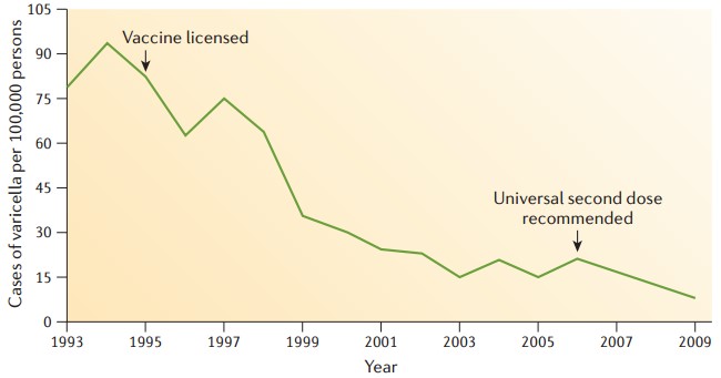 Figure 2. Epidemiological changes of VZV before and after varicella vaccination
(Source: Gershon AA, et al. 2015)
Acute VZV infection is diagnosed by detection of the virus. One of the preferred methods is the polymerase chain reaction (PCR) to detect the viral genome in cystic fluid, cerebrospinal fluid, tissue, bronchoalveolar lavage, EDTA blood, or amniotic fluid. A more specific point is its ability to test cerebrospinal fluid in cases of suspected acute central nervous system infections, as well as amniotic fluid in prenatal diagnosis following chickenpox during pregnancy. In immunosuppressed patients with herpes zoster, detection of VZV DNA in the blood may be useful to verify the potential risk of spreading the infection. Isolation and culture of VZV is time-consuming, requires extensive experience, does not have clinically relevant sensitivity, and is only suitable for blister fluids containing high viral loads. Immunofluorescent assays using monoclonal fluorescein-labeled antibodies can correctly identify viral isolates, and commercially available kits are available for direct qualitative detection, but have low sensitivity and specificity.
Table 1. Methods for the detection of VZV
| Principle | Method | Patient samples |
| Detection of viral DNA | Polymerase chain reaction (PCR) | Vesicle content/swab, in 1 ml physiological saline or viral transport medium |
| Basic diagnostics | Cerebrospinal fluid, tissue, bronchoalveolar lavage, EDTA blood, amniotic fluid | |
| Viral isolation | Viral growth in cell culture, detection by monoclonal antibody | Vesicle content in viral transport medium with special swab, tissue, bronchoalveolar lavage |
| Special diagnostics | Transport under cooling (2–8 °C) | |
| Virus detection | Immunofluorescence test using monoclonal antibody | Cell-rich vesicle content in viral transport medium with special swab, tissue |
| Reduced sensitivity and specificity | ||
| Discrimination between wild-type/vaccine strains, genotyping | PCR, restriction fragment length polymorphism analysis, sequencing | Vesicle content in viral transport medium with special swab, tissue, cerebrospinal fluid, viral isolate |
| Special diagnostics |
(Source: Sauerbrei A 2016)
Serologic VZV diagnosis is indicated when there is a need to determine the status of susceptible populations to initiate active or passive immunoprophylaxis. There is no need to determine antibody status after varicella vaccination in healthy children, adolescents, and adults, as opposed to immunization status modification, which is more recommended for immunodeficiency vaccinees and healthcare workers.
In the laboratory, the specific antibody to VZV was detected by ligand test or partial immunofluorescence test. No matter which test method is used, each result interpreted by the respective laboratory as positive for anti-VZV antibodies can be used as a criterion for immunity against chickenpox. Commercial test kits have different sensitivities, so very low antibody titers cannot be recognized. Therefore, high sensitive detection methods, such as special glycoprotein enzyme-linked immunosorbent assay (ELISA) or membrane antigen fluorescent antibody (FAMA) test, should be used to control the immune status and carry out vaccine research after varicella vaccination. In addition, the reason for the low sensitivity of many commercially available VZV immunoassays may also be related to the occurrence of cross-reactivity with other herpesviruses.
Table 2. Methods for the determination of VZV-specific antibodies
| Method | Remark |
| Ligand assay (ELISA, chemiluminescence immune assay, etc.) | Determination and differentiation of Ig classes (IgG, IgM, IgA) in serum, plasma, and cerebrospinal fluid, based on whole viral antigen of VZV-infected cell cultures or viral glycoproteins |
| Simple performance, commercially available, automated | |
| Basic diagnostics | |
| Indirect fluorescence antibody test (IFAT) | Determination and differentiation of Ig classes (IgG, IgM, IgA) in serum, plasma, and cerebrospinal fluid |
| Simple performance, commercially available, requires experience for evaluation | |
| Basic diagnostics | |
| Fluorescence antibody membrane antigen (FAMA) test | Determination of antibodies against VZV glycoproteins in serum, reference test for determination of immunity |
| Special diagnostics | |
| VZV IgG avidity (ELISA or IFAT) | Differentiation between primary infection (varicella) and recurrent infection after virus reactivation (zoster) |
| Special diagnostics | |
| Neutralization test | Determination of antibodies against VZV glycoproteins in serum, good correlation with FAMA test |
| Special diagnostics |
(source: Sauerbrei A. 2016)
Consequently, inhibiting NRF2 activity holds promise as a therapeutic strategy for patients with NRF2-addicted cancers, as it can disrupt the cancer cells' reliance on NRF2-mediated cytoprotection. On the other hand, it is worth noting that NRF2 activation in immune cells within the host can significantly suppress cancer cell growth. This suggests that NRF2 inducers might also have therapeutic potential for treating certain cancers.
In 1974, Takahashi and his colleagues developed a live attenuated varicella vaccine in Japan, they isolated VZV from a boy with varicella and attenuated the virus by continuously culturing 33 generations in human and guinea pig fibroblasts. The live attenuated vOka consists of a mixture of different VZV genotypes and is distinguished from wild-type Oka by 42 single nucleotide polymorphisms. A large collaborative trial in 1979 demonstrated a high degree of safety and protection against varicella with the vOka vaccine, which was licensed in the U.S. in 1995 and recommended for routine immunization of healthy U.S. children up to 1 year of age, and a two-dose schedule of vaccination was recommended by the CDC in 2007. In countries where varicella vaccination is not available, temporary immune protection can be obtained through passive immunization (injection of VZV-specific antibodies).
The successful development of a varicella vaccine paved the way for shingles vaccine research. According to the results of the Shingles Prevention Study (SPS), the vaccine needs to be more potent to enhance specific cell-mediated immunity in older adults with latent VZV infection. In addition, the data showed that the herpes zoster vaccine reduced the burden of disease by 61.1%, with patients experiencing less pain and discomfort. The vaccine also reduced the incidence of herpes zoster, as well as the negative impact of herpes zoster on patients' ability to perform activities of daily living and health-related quality of life.
References
| Target | Cat. No. | Product Name | Size | Species | Application | Detection Sample | |
| VZV | DEIA472 | VZV IgG ELISA Kit | 96T | Human | Qualitative, Semiquantitative, Quantitative | Serum, Plasma and Cerebrospinal fluid | Inquiry |
| DEIA473 | VZV IgG and IgG avidity ELISA Kit | 96T | Human | Qualitative | Serum | Inquiry | |
| DEIA387 | Varicella-Zoster Virus IgG ELISA Kit | 96T | Human | Quantitative and qualitative | Serum, plasma or cerebrospinal fluid | Inquiry | |
| DEIA388 | Varicella-Zoster Virus IgA ELISA Kit | 96T | Human | Quantitative and qualitative | Serum, plasma or cerebrospinal fluid | Inquiry | |
| DEIA389 | Varicella-Zoster Virus IgM ELISA Kit | 96T | Human | Quantitative and qualitative | Serum, plasma or cerebrospinal fluid | Inquiry | |
| DEIA500 | VZV IgM ELISA Kit | 96T | Human | Qualitative | Serum | Inquiry | |
| DEIA501 | VZV IgG ELISA Kit | 96T | Human | Qualitative | Serum | Inquiry | |
| DEIA4425 | Varicella zoster virus IgG ELISA Kit | 96T | Qualitative | Cerebrospinal fluid, serum | Inquiry | ||
| DEIA-JY2141 | Human Anti-Varicella Zoster Virus (gE) Antibody IgG ELISA Kit | 96T | Human | Qualitative | Serum | Inquiry | |
| DEIA-NS2307-3 | Varicella Zoster Virus Glycoprotein E (VZV gE) ELISA Kit | 96T | N/A | Quantitative | Vaccine samples | Inquiry | |
| DEIA-NS2307-4 | VZV-IgG(Varicella zoster virus-Immunoglobulin G) ELISA Kit | 96T | Human | Qualitative | Serum or plasma | Inquiry | |
| DEIA-NS2307-5 | Human Anti-Varicella Zoster Virus (VZV/chickenpox) IgM ELISA kit | 96T | Human | Quantitative | Serum or plasma | Inquiry | |
| DEIA-NS2307-7 | Mouse Anti-Varicella Zoster Virus (VZV/chickenpox) IgG ELISA kit | 96T | Mouse | Qualitative | Serum or plasma | Inquiry | |
| DEIA-NS2307-8 | Mouse Anti-Varicella Zoster Virus (VZV/chickenpox) IgM ELISA kit | 96T | Mouse | Qualitative | Serum or plasma | Inquiry | |
| DEIA-NS2307-9 | Monkey Anti-Varicella Zoster Virus (VZV/chickenpox) IgM ELISA kit | 96T | Monkey | Qualitative | Serum or plasma | Inquiry |
Loading ......
