
Inquiry Basket
 Loading ......
VZV is a double-stranded DNA alpha-herpesvirus of humans, with no known animal reservoir. Cell types particularly susceptible to infection are T lymphocytes, epithelial cells, and nerve ganglia. Primary infection results in chickenpox, and virus becomes latent in neurons during this infection and persists in the ganglia. Reactivation of the latent infection with age or immunosuppression (cellular immunity diminishes) causes shingles.
Creative Diagnostics provide high-quality VZV-related antigens and antibodies to support accurate research and diagnostic applications.
The VZV virion consists of three layers:
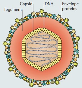 Figure 1. Virion formation and structure of VZV.
(Source: Zerboni L, et al. 2014)
When VZV particles reach the mucosal epithelial entry sites in humans, the virus begins to infect the host. It replicates locally and then spreads to the tonsils and other regional lymphoid tissues, where VZV infects T cells. The infected T cells transport the virus to the skin via the bloodstream. During primary infection, VZV goes into latency in sensory ganglia by two routes, either axonal transport to neuronal nuclei or transport via the bloodstream.
Following reactivation and return to viral replication, VZV moves via axons of nerves to the skin, where it causes shingles. Shingles is characterised by the vesicular rash that appears in the dermatome innervated by the affected ganglion. Lesions on the skin of both chickenpox and shingles lesions are known to have high titres of infectious virus and are the primary source of infection in uninfected hosts.
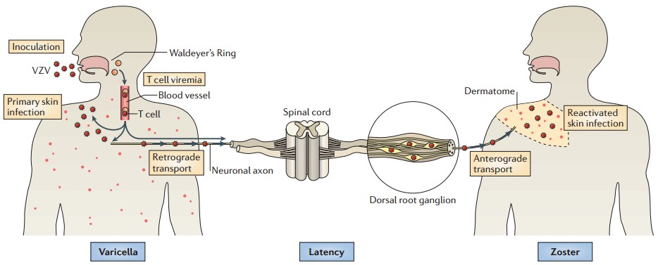 Figure 2. Model of the VZV life cycle.
(Source: Zerboni L, et al. 2014)
Varicella pneumonia is the most common complication of primary VZV infection in pregnancy. It has been reported to occur in 20% of pregnant women with chickenpox. Varicella pneumonia usually develops within 1 week of onset of the rash. Clinical features of varicella pneumonia include fever, dry cough, dyspnea, and mild hypoxia.
The fetal sequelae of maternal infection with VZV depend on the gestational age at the time of primary infection. First trimester infection does not appear to increase the risk of spontaneous abortion. First and second trimester infection can lead to fetal varicella syndrome (FVS) or congenital varicella syndrome (CVS) with the greatest risk of FVS between 13 and 20 weeks of gestation. The incidence of FVS decreases markedly after 20 weeks as the fetal immune system matures.
CVS
Neonatal Varicella
Table 1. Different approaches to managing varicella exposure in pregnant women. (Source: Ion A, et al. 2025)
| Approach | Indication | Contraindications | Advantages | Disadvantages |
| Screening and Vaccination (Pre-Pregnancy) | All women of childbearing age without documented immunity to VZV | Pregnancy (live attenuated vaccine contraindicated) | Prevents varicella during pregnancy; long-term immunity | Not applicable during pregnancy; requires pre-pregnancy planning |
| Anti-VZV Immunoglobulin (Post-Exposure) | Susceptible pregnant women (no history of chickenpox or VZV vaccination) within 10 days of exposure | Known immunity to VZV | Reduces risk of severe maternal varicella and congenital varicella syndrome (CVS); safe during pregnancy | Must be administered within a specific timeframe (72–96 h post-exposure for maximum efficacy) |
| Symptomatic Treatment + Oral Acyclovir | Pregnant women with active chickenpox rash or VZV exposure >10 days earlier | Severe renal impairment without adjustment or hypersensitivity to acyclovir | Reduces fever duration and symptom severity when started early (within 24 h of rash onset) | Limited efficacy if started late; primarily benefits the mother; potential adverse effects with prolonged use |
| Intravenous Acyclovir + Supportive Care | Severe maternal Varicella (E.G., Respiratory symptoms, complications, immunocompromised, or late pregnancy) | Hypersensitivity to acyclovir; cautious use in renal impairment | Effective in treating complications; reduces maternal mortality | Requires hospitalization; potential nephrotoxicity |
| Management During Last 4 Weeks of Pregnancy | Pregnant women with active varicella rash within 7 days before or after delivery | Nonspecific unless maternal contraindications exist | Reduces neonatal varicella severity when anti-VZV immunoglobulin is given to newborns | May not fully prevent neonatal varicella; requires close monitoring and additional treatment with IV acyclovir |
| Intravenous Acyclovir for Neonates | Neonates who develop varicella despite immunoglobulin prophylaxis | Hypersensitivity to acyclovir | Reduces severity and complications of neonatal varicella | Requires hospitalization; limited efficacy in preventing long-term complications if started late |
The first live attenuated VZV vaccine strain was obtained from wild virus, which was isolated from vesicular fluid taken from a chickenpox-infected infant. The wild virus was attenuated through repeated culturing on non-human cells. In 1974, the vaccine was first clinically used in Japan, and a group of children were successfully protected from chickenpox. This vaccine has since been approved in Japan, Korea, and the United States for clinical use. Many vaccines that were based on this strain are now available, such as monovalent vaccines against chickenpox and tetravalent vaccines for chickenpox, measles, mumps, and rubella, and many new vaccines are currently being developed.
Table 2. Pros and cons of live attenuated, inactivated or subunit vaccines against VZV. (Source: Freer G, et al. 2018)
| Vaccine | Pros | Cons |
| Live, attenuated |
|
|
| Heat-inactivated |
|
|
| Subunit |
|
|
References
Loading ......
