
Inquiry Basket
 Loading ......
SARS-CoV-2 is the virus responsible for COVID-19. It got its name from its connection to SARS-CoV but it differs greatly in how it spreads and the symptoms it causes. SARS-CoV-2 genetic material comprises positive-sense RNA made up of about 30kb while having an approximate GC content of 38%. The superior transmission of SARS-CoV-2 compared to SARS-CoV and MERS-CoV stems from its enhanced host adaptation abilities and its increased production of asymptomatic infections that promote human spread.
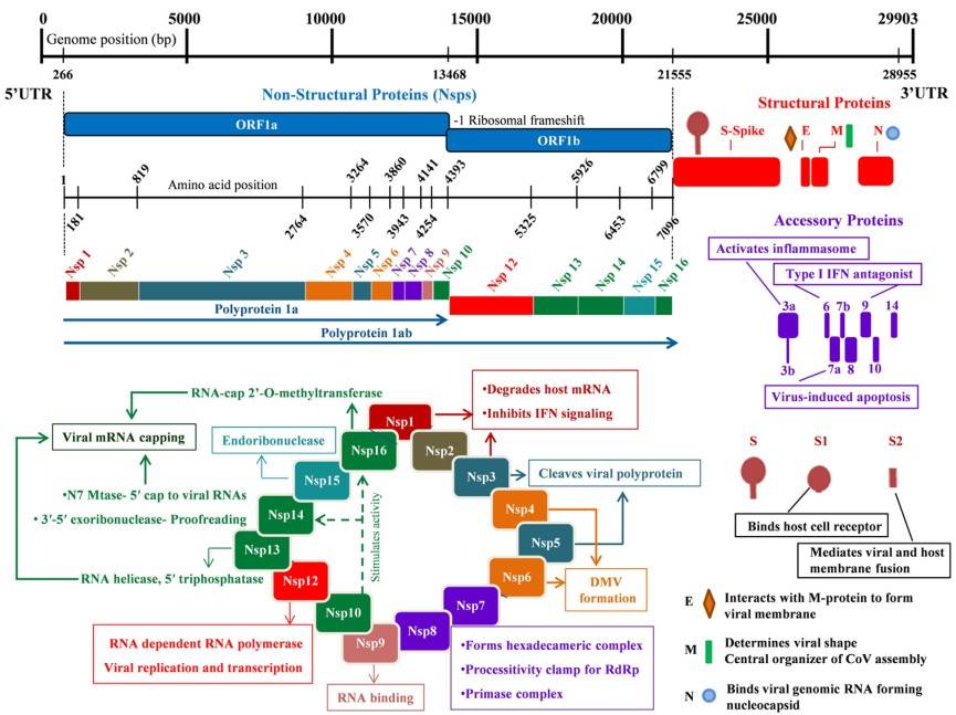 Figure 1. Genome organization of the SARS-CoV-2
(Source: Arya R, et al. 2021)
The SARS-CoV-2 genome features the replicase gene ORF1ab and structural protein genes S, E, M, N after its 5' UTR followed by accessory protein genes ORF3a and ORF6.
Viral entry, involving host cell membrane attachment and fusion, is mediated by the S protein. Coronaviral entry typically requires two spike (S) protein cleavage events: The first cleavage event occurs at the S1-S2 subunit junction while the second takes place at the S2' site inside the S2 subunit. Viral maturation in infected cells triggers cleavage of the polybasic motif at the S1-S2 boundary in SARS-CoV-2 but the S2' site cleavage occurs after the spike protein binds to ACE2 on target cells. The S1 subunit undergoes conformational changes upon ACE2 binding which exposes the S2' cleavage site located in the S2 subunit. Depending on the entry pathway, the S2' site is cleaved by distinct proteases. If target cells lack sufficient transmembrane protease serine 2 (TMPRSS2) or the virus-ACE2 complex does not encounter TMPRSS2, the complex is internalized via clathrin-mediated endocytosis into endolysosomes. In the presence of TMPRSS2, S2' cleavage occurs at the cell surface. In both pathways, S2' cleavage exposes the FP, and S1-S2 dissociation triggers large conformational rearrangements in the S2 subunit—particularly in HR1—driving FP insertion into the target membrane to initiate fusion. The merger of viral and cellular membranes forms a fusion pore through which viral RNA is released into the host cytoplasm.
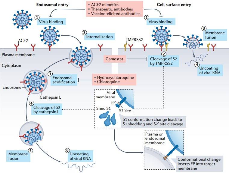 Figure 2. Two distinct SARS-CoV-2 entry pathways
(Source: Jackson CB, et al. 2022)
The S protein mediates viral entry through its structure which includes two subunits: S1 which contains the receptor-binding domain (RBD) and S2 which contains the fusion domain. By attaching to the host ACE2 receptor the RBD initiates structural alterations which reveal the S2 fusion peptide (FP) that facilitates virus-host membrane fusion. Cryo-electron microscopy studies have revealed two S protein conformations: The protein has two conformations where the RBD is hidden in the closed state and attached to ACE2 in the open state which becomes the main target for neutralizing antibodies. The S2 subunit exists in two different structural configurations which are the pre-fusion and post-fusion forms. In the pre-fusion state, the HR1 region forms a four-helix bundle as part of the S2 central helical stem, while HR2 remains disordered, and the FP adopts a short helix with a loop burying hydrophobic residues. Upon S1 subunit-receptor binding, the S2 subunit undergoes molecular maturation into the post-fusion conformation. First, S1 and S2 dissociate via proteolytic cleavage by host proteases, followed by the formation of a six-helix bundle coiled-coil structure through HR1/HR2 pairing in the trimeric S2. This transition brings the viral membrane closer to the host membrane, exposing the hydrophobic FP on either side of the HR helical bundle and rendering the region accessible for upstream proteolytic cleavage. Post-cleavage, FPs from each protomer in the trimeric assembly associate and fuse into the host membrane, enabling viral RNA release into the host cell.
The SARS-CoV-2 S protein includes a four-residue insertion (PRRA) at the S1/S2 boundary which results in the formation of a new Furin cleavage site. SARS-CoV-2 retains its ability to fuse with host cells after deletion of the extra cleavage site but this site likely contributes significantly to viral spread and infection rates.
 Figure 3. Distinct conformational states of the SARS-CoV-2 spike protein
(Source: Zhang J, et al. 2021)
As a key structural component, the N protein binds viral RNA using its NTD and generates dimers through its CTD for RNP complex formation. During virus particle assembly the N protein establishes interactions with other structural proteins which leads to encapsidation of the genome. Because the N protein generates a strong immune response and appears abundantly during infection it represents an ideal target for vaccine research.
SARS-CoV-2 N protein contains two domains that show a high degree of conservation. Disordered structures extend across both the N-terminal and C-terminal regions of the protein spanning residues 1–42 and 435–419. The disordered N-terminal area initiates liquid-liquid phase separation which allows viral genome packaging and prevents RNA interference by engaging with host proteins including hnRNPs. The serine/arginine-rich region controls the N protein's oligomerization state and subcellular localization which in turn influences viral replication efficiency.
The M protein is the most abundant structural protein and serves as the primary component of the viral envelope. The C-terminal domain of this protein interacts with the N protein to control viral particle assembly. The M protein consists of three major structural domains which include an N-terminal ectodomain and a transmembrane helical domain along with a C-terminal endo-domain. The transmembrane region of the protein demonstrates the ability to form dimers which stabilize the envelope structure using hydrophobic interfaces according to protein structure predictions.
The E protein is the smallest transmembrane structural protein, composed of 75 amino acids, and includes three domains: a hydrophilic ecto-domain at the N-terminal end and a hydrophilic endo-domain at the C-terminal end with a hydrophobic transmembrane domain located between them. The TMD reveals a pentameric helical bundle structure through NMR structural analysis which forms a narrow hydrophilic central pore that allows cation permeation and shares similarity to viroporins. The PDZ-binding motif at the C-terminal end of the E protein targets host proteins like PALS1 leading to disrupted cell junctions and tissue damage. Engineered coronaviruses without the E protein exhibit diminished viral titers and maturation defects and experience reduced replication rates which suggests it could serve as a vaccine candidate.
The nsps encoded by ORF1ab assemble into the RTC, which exhibits extensive intermolecular interactions. The viral replication machinery is anchored to convoluted membranes via the transmembrane proteins nsp3, nsp4, and nsp6, shielding dsRNAs from immune degradation.
Key components include:
Nsp3 is a multi-domain membrane-associated protein and the largest protein encoded by the coronavirus genome. It acts as a membrane-anchored scaffold, interacting with other nsps and host proteins to form the RTC. Nsp3 comprises the N-terminal Nsp3a domain, Macrodomain-X, SUD, PLpro, RBD, marker domain (MR), transmembrane domain (TM), and Y domain.
The PLpro enzyme processes viral polyprotein precursors pp1a and pp1ab through proteolytic cleavage at three exact positions resulting in the generation of nonstructural proteins Nsp1, Nsp2, and Nsp3. The PLpro enzyme of SARS-CoV-2 starts with an N-terminal ubiquitin-like domain that stands apart from its catalytic core domain while SARS-CoV PLpro remains unchanged. The catalytic core adopts an open right-handed architecture containing thumb, palm, and finger subdomains. PLpro functions as a galactose protease, with its active site located in a cleft between the thumb and palm subdomains. In unliganded PLpro structures, the flap adopts an open conformation. Upon inhibitor binding, the flap closes over the catalytic cleft entrance via an induced-fit mechanism, forming intermolecular interactions with the ligand. The loop can assume multiple conformations depending on the size and type of the inhibitor molecule.
3CLpro, which is also called Nsp 5 (3C-like protease), functions as a 33.8 kDa cysteine protease that processes the viral polyproteins pp1a (486 kDa) and pp1ab (790 kDa). The 3CLpro monomer comprises three domains: The 3CLpro protein consists of Domain I which spans residues 8–101, Domain II which spans residues 101–184, and Domain III which spans residues 201–303 along with a long connecting loop between Domains II and III which spans residues 185–200. The active site of 3CLpro emerges from the space between Domains I and II and consists of a Cys-His catalytic dyad made up of Cys145 and His41. The nsp5 protein from SARS-CoV-2 demonstrates 96% sequence homology compared to the SARS-CoV version of this protein. 3CLpro plays an essential role in the viral life cycle through its direct acceleration of nsp maturation. Research into its structure and catalytic mechanism has demonstrated that this protein represents a valuable target for the development of drugs against coronavirus.
The 932 amino acid long RNA-dependent RNA polymerase SARS-CoV-2 nsp12 includes a polymerase domain structured as a "right-hand grip" and an N-terminal domain. The finger domain of SARS-CoV-2 nsp12 includes three helices and a helix-loop-helix motif typical for RdRp along with two-stranded β-sheets as structural components while the palm domain contains two helices and a β-hairpin structure that holds catalytic aspartates for nucleotide transfer functions. The binding site for the nsp7-nsp8 heterodimer exists at the interaction site between the thumb and index finger subdomains while nsp12 and nsp7 maintain most of these contacts. The coronavirus family shows high conservation levels in core functional regions including the template entry site, template-primer exit site, nucleoside triphosphate (NTP) tunnels and the polymerase active site.
The addition of nsp7 and nsp8 cofactors significantly enhances nsp12 activity. Although other viral factors are required, the nsp12-nsp7-nsp8 complex is indispensable for nucleotide polymerization. Similar to the SARS-CoV polymerase complex, the nsp7-nsp8 heterodimer interacts above the thumb subdomain of nsp12. This interaction is primarily mediated by nsp7, while nsp8 exhibits limited contact with the polymerase subunit nsp12. Another nsp8 subunit engages the top region of the finger domain and forms additional interactions with the interface domain. However, the RNA synthesis efficiency of the SARS-CoV-2 nsp12-nsp7-nsp8 complex (~35%) is lower than that of the SARS-CoV counterpart due to alterations in the nsp8 subunit. Additionally, residue substitutions in SARS-CoV-2 nsps result in relatively lower thermal stability for both nsp8 and nsp12.
ORF3a functions to trigger cell death while enhancing viral pathogenicity and facilitating viral release. The activation of inflammasomes through ORF3a may trigger the cytokine storm effects observed in COVID-19 patients according to a group of scientists. The extrinsic apoptotic pathway activated by ORF3a begins with caspase-8 cleavage before proceeding to Bid cleavage which leads to cytochrome c release from mitochondria and culminates in apoptosome formation and caspase-9 activation. The reduced capability of SARS-CoV-2 to induce cell death in comparison to SARS-CoV may explain why these viruses cause different levels of disease severity. The pro-apoptotic activity together with membrane localization is lost through mutations at residues C130, C133, and Y160 which could affect viral pathogenicity. Blocking SARS-CoV ORF3a expression leads to decreased viral release and reduced disease severity.
ORF7b overlaps with SARS-CoV ORF7a on the same RNA segment. During SARS-CoV-2 infection in Vero cells the protein moves to the Golgi apparatus where it connects with internal virions and purified virus particles. The function of this protein remains unknown in both the SARS-CoV and SARS-CoV-2 viruses according to current scientific research.
References
| Target | Cat. No. | Product Name | Size | Species | Application | Detection Sample | |
| SARS-CoV-2 | DEIASL019 | SARS-CoV-2 IgG ELISA Kit | 96T | Qualitative | Serum | Inquiry | |
| DEIASL020 | SARS-CoV-2 IgM ELISA Kit | 96T | Human | Qualitative | Serum, plasma | Inquiry | |
| DEIASL024 | SARS-CoV-2 Total Antibody ELISA | 96T | Human | Qualitative | Serum, plasma | Inquiry | |
| DEIASL055 | SARS-CoV-2 Neutralizing Antibody Assay Kit | 96T | Qualitative | Serum, plasma | Inquiry | ||
| SARS-CoV-2 Spike glycoprotein | DEIASL411 | SARS-CoV-2 Spike RBD ELISA Kit | 96T | Quantitative | Cell culture supernates, plasma | Inquiry | |
| DEIASL193 | SARS-CoV-2 S1 IgG Titer ELISA Kit | 96T | Quantitative | Serum, plasma | Inquiry | ||
| DEIASL018 | SARS-CoV-2 Spike glycoprotein ELISA Kit | 96T | Quantitative | Cell culture supernates, plasma | Inquiry | ||
| SARS-CoV-2 NP | DEIASL194 | SARS-CoV-2 Nucleocapsid Protein IgG Titer ELISA Kit | 96T | Quantitative | Serum, plasma | Inquiry | |
| DEIASL017 | SARS-CoV-2 N ELISA Kit | 96T | Human | Quantitative | Serum, plasma | Inquiry |
Loading ......
