
Inquiry Basket
 Loading ......
In 1928 the bacteriologist Alexander Fleming discovered that Penicillium notatum can secrete an antimicrobial molecule that can inhibit bacterial growth in vitro and named it penicillin. The discovery of penicillin changed the course of history and set the stage for the antibiotic era and the subsequent development of other, more effective antibiotics. The original molecule extracted from P. notatum was originally called benzylpenicillin (penicillin G), and today penicillin refers to different molecules that has the structure of a β-lactam and has the same antimicrobial activity as penicillin G. Penicillin has saved millions of lives since its discovery, including children, pregnant women, patients with sepsis, meningitis or endocarditis, and other life-threatening infections, and today penicillin G remains the only recommended treatment for preventing mother-to-child transmission of syphilis.
To address the problem of penicillin resistance, pharmacologic research has begun to develop a new generation of penicillins. The second generation of penicillins are semisynthetic β-lactamase-resistant penicillins such as oxacillin, methicillin, and dicloxacillin, which resist the enzyme penicillinase in staphylococci, and are therefore also known as antistaphylococcal penicillins. The spectrum of activity of this class of antibiotics is relatively narrow, and in order to provide wider coverage of Gram-negative bacteria, a third generation of broad-spectrum penicillins (also known as aminopenicillins) was introduced, with amoxicillin and ampicillin being the main representatives of this class of penicillins. Third-generation penicillins are more effective against a wider range of Gram-negative bacteria because they are more stable to penicillinase. The last generation of penicillins, carboxypenicillins and ureidopenicillins, further extended the range of penicillin action against Gram-negative bacteria and showed potent activity against Pseudomonas aeruginosa.
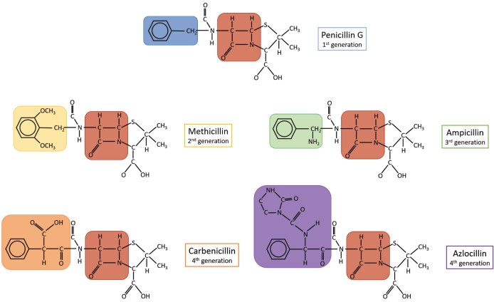 Figure 1. Chemical structures of different classes of penicillins
(Source: Lobanovska M, et al. 2017)
The cell wall is the basic structural element of most bacteria and consists of peptidoglycan (PGN), whose main function is to maintain the integrity and shape of the cell and to prevent macromolecules from penetrating the cell. Peptidoglycan is located outside the cytoplasmic membrane and consists of alternating chains of N-acetylglucosamine (GlcNAc) and N-acetylmuramic acid (MurNAc) residues, which are covalently cross-linked by short peptides. During growth and division, PGN is synthesized and remodeled. PGN is characterized by its reticular conformation, which is derived primarily from peptide cross-linking. These connections are formed in the presence of specific enzymes called transpeptidases or penicillin-binding proteins (PBPs). Penicillin, like other β-lactam antibiotics, contains a four-membered β-lactam ring that inhibits transpeptidases. By mimicking the last two D-alanine residues of the peptide, penicillin is able to irreversibly bind to the active site of transpeptidases, preventing the enzyme from cross-linking the peptidoglycan chains. Since penicillin prevents the formation of new PGNs, the existing PGNs in the bacterial cell wall do not provide resistance to osmotic stress, and the cell is therefore susceptible to lysis. It is worth noting that since eukaryotic cells lack both PGN and the enzyme responsible for PGN synthesis, penicillin targets only bacteria and does not affect eukaryotic cells.
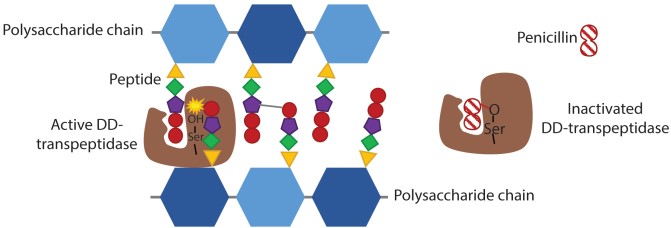 Figure 2. Schematic representation of the mechanism of penicillin action
(Source: Lobanovska M, et al. 2017)
Penicillins can be categorized on the basis of chemical substitutions of residues on the β-lactam ring, which give them different activities. For example, penicillin G is more effective against Gram-positive bacteria, especially cocci and bacilli, but less effective against Gram-negative bacteria. This failure to act against Gram-negative bacteria is also present in other penicillin antibiotics, mainly because Gram-negative bacteria contain an outer membrane that prevents penicillin penetration. Secondly some Gram-negative bacteria have acquired specific genes encoding penicillinases that hydrolyze the β-lactam ring to inactivate penicillin.
Penicillin is the most common drug allergy found in medical records, with prevalence rates varying across regions and populations treated. Penicillin has been the most common cause of fatal and nonfatal allergic reactions to drugs in the United States and the United Kingdom. Benign skin reactions such as urticaria and delayed maculopapular rash are the most common types of reactions. The lowest incidence of anaphylaxis is associated with oral penicillins, and aminopenicillins are one of the drugs at high risk for causing a benign delayed rash, usually in the context of an acute EBV infection. In addition, aminopenicillins are considered the most common cause of acute generalized exanthematous pustulosis (AGEP). The administration of penicillins may also cause severe skin reactions in some patients, such as drug reactions with eosinophilia and systemic symptoms (DRESS).
Penicillin is a semi-antigen, and the β-lactam ring binds to lysine residues in serum proteins, and when bound to the polylysine substrate, it produces the major antigenic determinant, penicillamide polylysine. Clinical immune-mediated responses associated with penicillins are primarily antibody-mediated or T-cell-mediated. Immune responses mediated by antibodies can be divided into three categories, of which type I hypersensitivity is mediated by IgE. Dendritic cells bind and internalize penicillin-binding proteins for presentation to naive CD4+ T cells (type 0 helper T cells). In the presence of interleukin-4 (IL-4), naive T cells develop as penicillin-specific type 2 helper T (Th2) cells. The latter produce IL-4 and IL-13, which induce B cells to differentiate into penicillin-specific IgE-secreting plasma cells. IgE binds to Fc epsilon receptors on the surface of basophils and mast cells, and on re-exposure, polyvalent penicillin cross-links the Fc epsilon receptor bound to the IgE antibody, inducing degranulation of mast cells and the release of soluble inflammatory mediators such as histamine, prostaglandins, and leukotrienes, which leads to the clinical manifestations of anaphylaxis. In type II reactions, antibodies or immune complexes target the cell membrane structure of red blood cells, white blood cells, or platelets, leading to cell destruction or sequestration and ultimately hemolytic anemia and thrombocytopenia. In type III reactions, antibodies formed within 4 to 10 days react with the penicillin protein carrier to form soluble immune complexes. Activation and deposition of complement in small vessels leads to recruitment of neutrophils by Fc-IgG receptors, which releases protein hydrolases leading to tissue damage and localized vascular inflammation such as small vessel (hypersensitivity) vasculitis and serum sickness.
 Figure 3. Antibody-mediated mechanisms of adverse reactions to penicillins
(Source: Castells M, et al. 2019)
T-cell-mediated reactions usually occur more than 6 hours after penicillin administration or during treatment with multiple exposures to penicillin. Delayed responses are usually associated with models involving non-covalent binding, such as models of pharmacological interactions or alterations in HLA peptide presentation specificity. Antigen-presenting cells process drug-modified peptides and present them to the antigen-binding groove of the HLA for recognition by T-cell receptors (TCRs) on CD4+ or CD8+ T-cells, leading to T-cell activation and release of cytokines and chemokines. Common phenotypes include reactions within 1 to 6 hours after exposure (urticaria and anaphylactic reactions), and reactions occurring more than 6 hours after a single or multiple administrations (macular papules). Delayed T-cell-mediated responses with systemic involvement include severe cutaneous reactions (SJS-TEN, DRESS, and AGEP).
 Figure 4. T cell mediated mechanisms of adverse reactions to penicillins
(Source: Castells M, et al. 2019)
A thorough history evaluation of patients reporting penicillin allergy is necessary, and in some cases, allergists are readily available to perform penicillin allergy evaluations and to assist in the development of protocols, pathways, and referral processes to maximize the safety and efficiency of these evaluations. However, most patients reporting penicillin allergy may be evaluated by a non-specialist. Based on allergy history, patients can be risk-stratified, which helps clinicians decide whether a penicillin skin test or drug challenge is appropriate.
Table 1. Risk Stratification for Penicillin Allergy Evaluation
| Low Risk | Medium Risk | High Risk | |
| History | Isolated reactions that are unlikely allergic Pruritus without rash Remote (>10 y) unknown reactions without features of IgE Family history of penicillin allergy | Urticaria or other pruritic rashes Reactions with features of IgE but not anaphylaxis | Anaphylactic symptoms Positive skin testing Recurrent reactions Reactions to multiple β-lactam antibiotics |
| Action | Prescribe amoxicillin course or perform a direct amoxicillin challenge under observation | Skin test followed by amoxicillin challenge under observation if the skin test is negative. Consider allergy/immunology referral | Allergy/immunology referral or desensitization. |
(Source: Shenoy ES, et al. 2019)
Direct oral challenge with amoxicillin (DOCA) under supervision without allergy testing was recently reported to be safe in low-risk patients. Amoxicillin contains potentially antigenic side chains and accounts for the majority of penicillin antibiotic use, amoxicillin tolerance not only precludes selective amoxicillin allergy due to side-chain reactivity, but also ensures tolerance to the broader penicillin family.
Single-step or two/three-step DOCA with a cumulative dose of 500mg under the supervision of a physician. In patients with an unknown history or who present with non-immediate adverse reactions, a further course of 3-5 days may be given to rule out immediate and non-immediate hypersensitivity reactions. Patients were selected for DOCA based on a guideline-based risk stratification process, and those stratified as high risk were referred to an allergy specialist for further evaluation.
References
| Target | Cat. No. | Product Name | Size | Species | Application | Detection Sample | |
| PCN | DEIABL-QB24 | Penicillin ELISA Kit | 96T | N/A | Quantitative | Animal tissue (muscle, liver, shrimp and fish), milk etc | Inquiry |
| DEIABL-QB25 | Benzyl penicillin ELISA Kit | 96T | Benzyl penicillin | Quantitative and Qualitative | N/A | Inquiry | |
| Amoxicillin | DEIABL-QB49 | Amoxicillin ELISA Kit | 96T | Quantitative | Chicken, swine meat, milk | Inquiry | |
| Dicloxacillin | DEIABL-QB27 | Dicloxacillin ELISA Kit | 96T | Quantitative | Honey, tissue | Inquiry | |
| Oxacillin | DEIABL-QB30 | Oxacillin ELISA Kit | 96T | Quantitative | Tissue, milk | Inquiry | |
| Ampicillin | DEIA040 | Ampicillin ELISA Kit | 96T | N/A | Quantitative | Animal tissues, milk, honey | Inquiry |
Loading ......
