
Inquiry Basket
 Loading ......
LM stands out as a lethal foodborne pathogen that occurs frequently in contaminated foods. The natural environment hosts this organism which also occurs in poultry meat products and dairy items. Pregnant women infected with LM can develop severe invasive infections that result in miscarriage and stillbirth together with disseminated fetal infections. The bacterium LM appears as a Gram-positive short rod that requires minimal nutrients. The categorization of LM into 13 serotypes relies on O and H antigens, but more than 95% of isolates fall under serotypes 1/2a, 1/2b, 1/2c, and 4b. Serotypes 1/2a and 1/2b are food isolates but serotype 4b isolates approximately half of which comes from clinical patients. Using phenotypic characteristics and genotyping methods, LM is divided into four evolutionary lineages. The primary sources are lineages I and II while lineage III occurs less frequently and lineage IV shows rarity. Lineage I comprises six serotypes including 1/2b, 4b, 3b, 4e, 4d, and 7 while lineage II consists of four serotypes which are 1/2a, 1/2c, 3a, and 3c and lineages III and IV share serotypes 4b, 4a, and 4c.
 Figure 1. Three Listeria lineages
(Source: Radoshevich L, et al. 2018)
LM uses virulence factors as essential invasion tools and its ability to cause disease depends on four virulence island genes (LIPI-1, LIPI-2, LIPI-3, LIPI-4) and the stress survival island 1 (SSI-1) gene. The LIPI-1 virulence island contains important intracellular survival genes which include prfA, plcA, hly, mpl, actA, and plcB. The prfA gene produces PrfA which acts as a transcriptional activator to control the transcription process of over 140 genes within LIPI-1. PrfA produced from the prfA gene functions as a transcriptional activator to regulate transcription of more than 140 genes that are part of LIPI-1. The plcA gene produces PI-PLC while the plcB gene produces PC-PLC and both proteins function alongside listeriolysin O (LLO) to allow Listeria monocytogenes to enter the cytoplasm through the dissolution of single-membrane vacuoles by PI-PLC. Through the formation of actin filaments ActA collaborates with PC-PLC to enhance cell-to-cell movement which increases Listeria monocytogenes attachment to cell membranes. LLO, encoded by the hly gene, exerts its lytic activity in epithelial cells, allowing Listeria monocytogenes to disrupt and invade host secondary phagosomes and facilitates its translocation to the host cytoplasm. In the bloodstream, LLO helps Listeria monocytogenes escape phagocytosis by immune cells and proliferate in the cytoplasm. hly deletion mutants show significantly reduced ability to invade epithelial cells and markedly decreased intracellular proliferation, leading to a sharp decline in bacterial virulence. LLO can also alter host gene expression, helping to weaken the immune response against Listeria monocytogenes infection. mpl encodes a zinc metalloprotease that is a key enzyme for the maturation of pro-plcB. prfA and hly in LIPI-1 have been widely used as target genes for detecting Listeria monocytogenes, applied in various food safety tests and virulence assessment of isolates. The virulence island LIPI-1 increases the invasiveness of Listeria monocytogenes, and its virulence is closely related to the presence of surface proteins InlA, InlB, and LLO.
The virulence genes inlA through inlH that LIPI-2 contains play roles in Listeria monocytogenes adhesion and invasion functions. The Listeria monocytogenes surface proteins InlA and InlB facilitate host cell internalization which helps the bacteria form biofilms to increase survival in food processing settings. The proteins Internalins (InlA-H) produced by genes inlA to inlH enable host cells to engulf Listeria monocytogenes during infection. InlA functions as a key factor enabling Listeria monocytogenes to penetrate both the intestinal epithelium and the placental barrier. The LRR domain of InlA binds to the actin cytoskeleton and disrupts E-cadherin functionality while triggering cortical actin polymerization and plasma membrane changes which enable cell invasion and passage through intestinal and fetomaternal barriers. InlB helps Listeria monocytogenes evade immune surveillance and invade liver cells. InlC is produced when Listeria monocytogenes enters the host cell cytoplasm and promotes the spread of secretory internalins between polarized epithelial cells. InlC binds to IKKα, preventing the phosphorylation and degradation of IκB, thereby avoiding the translocation of nuclear transcription factor κB (NF-κB), inhibiting gene transcription, and blocking NF-κB activation in macrophages. InlC can weaken the effect of inflammatory cytokines and hinder neutrophil aggregation at the infection site.
LIPI-3 mainly encodes Listeriolysin S (LLS), which can alter the host gut microbiota and cause cell lysis, thereby facilitating Listeria monocytogenes to cross the intestinal-blood barrier. LIPI-4 belongs to the cellobiose-family phosphotransferase system and is responsible for the neurotropic and placental infections of Listeria monocytogenes. LIPI-4 plays a role in crossing the blood-brain barrier and enhances the neurotoxicity and placental toxicity of Listeria monocytogenes. The stress survival islands include SSI-1 and SSI-2. SSI-1 mainly contributes to Listeria monocytogenes tolerance to low pH and high salt environments, while SSI-2 is involved in alkaline and oxidative stress tolerance and is found only in serotype 1/2a.
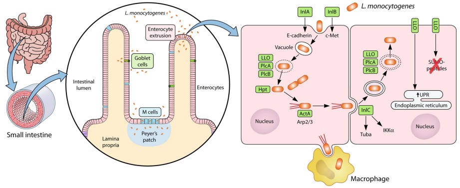 Figure 2. Pathogenesis of L. monocytogenes infection
(Source: Koopmans MM, et al. 2023)
The hly gene encodes LLO which functions as a member of the cholesterol-dependent cytolysins (CDC) family known for creating membrane pores. The toxin enables bacteria to disrupt phagosomes which allows them to enter the cytosol needed for proliferation within the cytoplasm. The protein LLO enables Listeria monocytogenes to break free from internalized host cell vacuoles by forming pores in the membrane. LLO functions as the primary virulence factor for Listeria monocytogenes by driving both primary and secondary phagosome escape mechanisms. Crystallographic studies show that the LLO monomer structure is highly similar to other CDC monomers and consists of four structural domains (D1 to D4). The primary structure of the monomer consists of domains D1, D2, and D3. The 25-amino acid signal peptide sequence and the PEST sequence region made of proline, glutamic acid, serine, and threonine in D1 enable Listeria monocytogenes to escape the phagosome. The D4 domain contains a highly conserved sequence and three loop structures, which are necessary for LLO monomer recognition of cholesterol and membrane binding.
During infection LM becomes trapped inside phagosomes within mammalian cells as an intracellular parasite since this enclosure impedes its ability to replicate. The mild acidity inside phagosomes triggers the production and release of LLO which enables LM to leave the phagosome and move into the cytoplasm. Research demonstrates that LM's ability to escape the phagosome and replicate ceases when LLO is absent or neutralized by specific antibodies. When LLO is present pathogens that normally stay within phagosomes like Escherichia coli manage to break through phagosome barriers to access the cytoplasm. LLO-generated membrane pores lead to a short-term rise in cytosolic calcium levels which triggers several cell signaling mechanisms including MAPK activation and PI signaling as well as NF-κB and cytokine release like IL-6, IL-8, and GM-CSF. Research shows that LM infection triggers multiple types of apoptosis. Scientific studies demonstrate that LLO triggers quick apoptosis of lymphocytes in both laboratory settings and living organisms.
The pathogenic nature of the intracellular parasite Listeria monocytogenes arises from multiple virulence genes working together. Listeria monocytogenes enters host cells through phagocytosis by gastrointestinal epithelial cells which allows it to penetrate the mucosal barrier and gain access to the bloodstream where it circulates to disseminate through the body. Among all human organs, the placenta and nervous system display the highest vulnerability to infection. The infection process includes internalization, escape, phagocytosis, actin filament aggregation, and intercellular spread, with each step requiring specific factors.
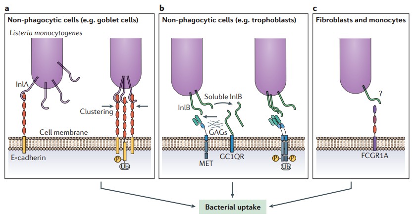 Figure 3. Entry of Listeria monocytogenes into cells
(Source: Radoshevich L, et al. 2018)
Listeria monocytogenes invades macrophages and epithelial cells directly to grow and multiply within them. The infection process of LM in macrophages involves several mechanisms including intracellular adaptation followed by cellular immune modulation and cell structure disruption while avoiding autophagy and extracellular traps (ETs). To survive within macrophages and resist nutritional immunity, Listeria monocytogenes enhances its adaptability to the intracellular environment by regulating the expression of virulence factors while scavenging nutrients inside the cell. LM proliferates in the host cytoplasm using hexose phosphate (HP), and HP transport proteins are key virulence factors for bacterial acquisition of HP, with their expression positively regulated by the transcription factor PrfA. During LM infection of macrophages, PrfA promotes bacterial intracellular survival and induces the secretion of bacterial foldase PrsA2 and protease HtrA. PrsA2 helps to fold secreted bacterial proteins and maintains their stability thereby increasing LM virulence and adaptability inside host cells. The expression of the surface protein ActA is regulated by this mechanism to enable bacterial movement within host cells' cytoplasm through actin polymerization control and facilitates persistent infection of neighboring cells while avoiding immune detection.
LM can evade macrophage phagocytosis by suppressing macrophage immune responses. LM carries two chitinase-encoding genes (chiA and chiB) and one chitin-binding protein-encoding gene, whose expression enhances bacterial virulence. Studies have found that Listeria monocytogenes chitinase ChiA enhances bacterial survival in host tissues by downregulating iNOS expression. This function shifts the host immune response from Th1-type inflammation to Th2-type inflammation and assists the bacteria in resisting host clearance by suppressing immunity. The internalin InlC promotes bacterial movement and spread between cells by stimulating the expression of membrane protrusion proteins. The bacteria produce large amounts of InlC to help resist macrophage immunity; InlC interacts with IKKα to reduce IκB degradation, thereby weakening the host immune response by inhibiting the NF-κB signaling pathway. The hemolysin LLO forms pores on the phagosome membrane and cooperates with bacterial Plc and metalloprotease Mpl to disrupt the phagosome. After phagosome rupture, bacteria escape into the macrophage cytoplasm. LLO and Plc inflict physical and biochemical damage to the phagosome membrane while simultaneously altering its maturation process to enable bacterial escape into the cytoplasm. Research shows the LM uses ActA or InlK to recruit the Arp2/3 complex and Ena/vasodilator-stimulated phosphoprotein (VASP) to the bacterial surface for macrophage autophagy evasion.
Listeria monocytogenes establishes host colonization through genetic and environmental strategies to form biofilms and create distinct infection pathways that differ from those of other intestinal pathogens. During bacterial growth cells establish a self-regulating biofilm community which adapts to environmental conditions by producing sticky extracellular polymeric substances (EPS) that bind to surfaces and multiply. The biofilm structure contains bacterial cells together with polysaccharides nucleic acids and proteins. Biofilms formed by LM on food contact surfaces are a key factor in the persistent contamination of food by pathogens. EPS provides mechanical stability to the biofilm, regulates its adhesion to surfaces, and forms a cohesive three-dimensional polymer network that connects, immobilizes, and traps biofilm cells. Bacteria embedded in the polymer matrix can avoid exposure to harsh external conditions. Once LM enters the food processing chain, planktonic bacteria detach from the biofilm in the later stages of biofilm formation and disperse into the surrounding environment, initiating a new round of biofilm formation. This leads to persistent cross-contamination and increases its spread throughout the food processing process.
The serotype of LM affects its biofilm formation ability. Different serotypes have varying pathogenicity, with serotypes 1/2a, 1/2b, and 4b commonly associated with listeriosis outbreaks. Isolates of serotypes 1/2a and 1/2c form biofilms more easily than those of 1/2b and 4b. Intracellular genes related to LM biofilms mainly include prfA, actA, flaA, and inlA. PrfA is a key positive regulator of gene expression and a biofilm development initiator. Low extracellular levels of PrfA allow bacteria to remain at the initial adhesion site and promote invasion and biofilm maturation after adhesion. ActA mediates LM aggregation and intercellular interactions, promoting clustering and biofilm formation. The presence of internalins, stress survival islands, and erythromycin resistance gene is associated with biofilm formation ability. Intercellular gene regulation related to LM biofilms includes agr, dlt, fliI, InlA, BapL, and PlcA. The accessory gene regulator (agr) system regulates biofilm formation and virulence. The agr locus consists of four genes: agrB, agrD, agrC, and agrA. Especially, agrB, agrC, and agrD are crucial during the initial adhesion stage of LM biofilm production. Additionally, the expression levels of the agr locus and prfA gene are strongly influenced by time and temperature.
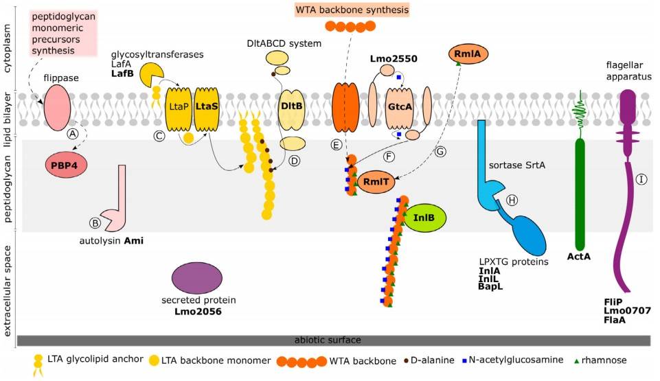 Figure 4. A schematic diagram of the Listeria monocytogenes surfactome and its structures affecting biofilm formation on abiotic surface
(Source: Janež N, et al. 2021)
References
| Target | Cat. No. | Product Name | Expression System | Tag/Conjugate | Application | |
| L. monocytogenes | DAGP0192 | Recombinant Listeria monocytogenes p60 [His] | E. coli | His | ELISA, LF | Inquiry |
| DAG4326 | Recombinant L. monocytogenes Internalin [His] | E. coli | His | N/A | Inquiry | |
| DAG-P2903 | Active Listeriolysin (aa 60 - 529) | E. coli | Unconjugated | SDS-PAGE | Inquiry | |
| DAG-P2785 | Active Listeriolysin (aa 26 - 529) | E. coli | Unconjugated | SDS-PAGE | Inquiry | |
| L. monocytogenes FliC | DAG-H10322 | L. monocytogenes Flagellin [His] | E. coli | His | N/A | Inquiry |
Loading ......
