
Inquiry Basket
 Loading ......
The etiological agent responsible for tuberculosis is Mycobacterium tuberculosis. Complex lipids including mycolic acids in its cell wall make it resistant to desiccation and extreme pH environments and enable acid-fast staining characteristics. Mycobacterium tuberculosis endures for several months when dried in sputum yet succumbs to moist heat at 63°C applied for 15 minutes. MTB is an obligate aerobe that grows slowly on Löwenstein-Jensen medium, forming characteristic cauliflower-like colonies in 2 to 4 weeks. BCG is an attenuated strain derived from MTB through continuous passage, losing some virulence genes, but its protective efficacy is significantly limited by geographic variation. MTB predominantly inhabits immune system cells in the human body which normally defend against or eliminate most bacterial infections. MTB only survives naturally within human beings who act as its host and reservoir. The bacterium MTB has developed specific physiological adaptations enabling its survival inside immune cells while establishing a mutual relationship between the cells' normal functions and its pathogenic activities.
MTB is transmitted through airborne droplets. The bacteria establish a long-term asymptomatic latent state that persists for decades or throughout the host's life after infection. A small percentage of people with latent tuberculosis infection experience reactivation that transforms the disease into a symptomatic and infectious stage. During latent infection, both immunocompetent individuals and those with compromised immune systems carry bacteria populations that exist in a non-replicating or slowly replicating state acting as potential sources for disease reactivation and transmission and therapeutic relapse. Once in the lungs, MTB is engulfed by immune cells and briefly replicates, then spreads via lymph and blood to other parts of the lungs. The immune system forms granulomas to contain and control MTB, keeping it in a dormant state.
MTB infects a range of host cells both immune and non-immune, but the macrophage stands as its primary host cell. The bacteria primarily reside in the phagosomes inside macrophages, separated from other microbes. MTB encounters dynamic host defenses inside the phagosome which include environmental acidification and the production of reactive oxygen and nitrogen intermediates along with antimicrobial peptides. MTB now possesses the capability to adapt to various biochemical stresses and partially bypass these defenses while entering the cytoplasm. MTB demonstrates the capability to survive for extended periods inside granulomas yet these structures can deteriorate which triggers cell death and caseous necrosis that exposes MTB to lipid-rich hypoxic extracellular surroundings. MTB completes its life cycle by developing infectious aerosols in the airways which then transmit to new hosts. Additionally, studies have found that MTB exists in multiple states within the host, some dormant and some difficult to detect by conventional culture methods.
 Figure 1. The mycobacterial circle of life
(Source: Ehrt S, et al. 2018)
The pathogenicity of MTB depends on the synergistic action of secreted proteins and cell wall antigens.
The Ag85 complex (Ag85A/B/C) is the most extensively studied virulence factor. This protein is secreted by both MTB and BCG cultures and exhibits a molecular weight of approximately 30 kDa. Mycobacteria share the Ag85 antigen complex which includes three components Ag85A, Ag85B, and Ag85C encoded by genes Rv3804c, Rv1886c and Rv0129c. The Ag85 complex components are secreted in a 2:3:1 ratio and they trigger both humoral and cellular immune responses. The Ag85 complex functions as a mycolyl transferase that attaches mycolyl groups to trehalose resulting in arabinogalactan-bound mycolic acid esters along with trehalose dimycolate which are fundamental in forming both inner and outer layers of the mycobacterial cell wall. The Ag85 complex attaches to human fibronectin at the cell surface and plays an essential role in MTB survival, pathogenicity and drug resistance.
MTB secretes Ag85B as its principal antigen which demonstrates strong immunogenic capabilities alongside serving as the primary target for macrophage phagocytosis. This major immunogen triggers Th1-type immune responses (IFN-γ, TNF-α) and CD8+ T cell cytotoxicity. Because of its properties scientists incorporated Ag85B in the development of recombinant BCG and viral vector vaccines. However, clinical trials have shown that its protective effect is significantly influenced by adjuvants and the genetic background of the population.
ESAT-6 protein is an intracellular protein and an early secreted protein isolated from Mycobacterium tuberculosis early culture filtrate (ST-CF). It is encoded by the Rv3875 gene in the R1 region that is deleted in BCG, making it one of the best antigens for distinguishing pathogenic MTB, non-pathogenic MTB, and BCG. ESAT-6 is an important T cell antigen that can recognize immunoactive T lymphocytes in the early stages of infection, inducing host-specific CD4+ T cell and CD8+ CTL proliferation and massive interferon-gamma production, thus providing immune protection. ESAT-6 also possesses B lymphocyte epitopes and can stimulate the production of specific immunoglobulins. However, since there is no ESAT-6 signal peptide sequence on the Mycobacterium tuberculosis cell membrane, the mechanism of its secretion to the extracellular space remains unclear.
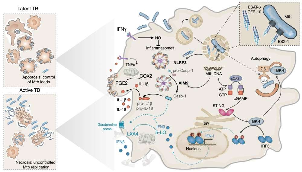 Figure 2. Effects of ESAT-6 on the outcome of MTB infection in host macrophages
(Source: Anes E, et al. 2023)
CFP10 is encoded by the Rv3874 gene in the R1 region deleted in BCG and is another early secreted protein isolated from Mycobacterium tuberculosis early culture filtrate. It exists only in pathogenic MTB and is absent in non-pathogenic mycobacteria such as BCG. The genes encoding ESAT-6 and CFP10 are adjacent and translated from the same operon, forming a stable 1:1 complex to exert their function. The widely used clinical IGRA test utilizes the ability of ESAT-6 and CFP10 expressed by MTB to stimulate peripheral blood mononuclear cells (PBMCs) from tuberculosis patients to secrete large amounts of interferon-gamma. After blood collection, interferon-gamma levels are measured within 12 to 18 hours. Compared to the tuberculin skin test, IGRA has higher sensitivity and specificity and is not affected by BCG vaccination or most non-pathogenic mycobacteria.
LAM is present in the cell wall of Mycobacterium tuberculosis and has various biological activities that can alter the host immune response. In patients with active tuberculosis, LAM is released from the bacteria and exerts immunoregulatory effects. It enters the bloodstream from tuberculosis lesions in the lungs or other affected sites and can relatively easily pass through the glomerular basement membrane, eventually being excreted in the urine. Compared to sputum, detecting LAM in urine is more convenient, which expands the application of rapid tuberculosis diagnosis.
LAM is chemically a lipopolysaccharide with thermal stability. LAM consists of four parts: a phosphatidylinositol anchor, a lipophilic mannan core, arabinofuranose branched side chains, and mannose cap motifs that modify the terminal side chains. Upon detection of MTB by host innate immune cells LAM functions as a PAMP which is identified by Toll-like receptors and C-type lectin receptors on macrophage and dendritic cell membranes to facilitate phagocytosis and trigger local immune responses. LAM demonstrates substantial immunosuppressive capabilities by blocking pro-inflammatory cytokine production and disrupting both phagosome maturation and macrophage apoptosis as well as impeding autophagy. These properties not only reduce the bactericidal capacity of phagocytes but also transform the interior of phagocytes into a safe sanctuary for self-replication, which may be an important reason for Mycobacterium tuberculosis to establish latent infection.
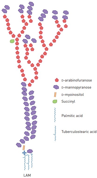 Figure 3. Schematic rendition of the structure of Mycobacterium spp. LAM
(Source: Correia-Neves M, et al. 2019)
MPT64, also known as MPB64, is a secreted protein of MTB encoded by the Rv1980c gene in the RD2 region, with a molecular weight of 24 kDa, accounting for 8% of the total secreted proteins of MTB. The BCG strain lost the MPT gene encoding this protein during passage. MPT64 is one of the most important Mycobacterium tuberculosis antigens in human-specific T cell responses. After MTB infects macrophages, MPT64 impairs the endoplasmic reticulum-mediated unfolded protein response in macrophages to facilitate long-term replication of MTB. MPT64 has good sensitivity, specificity, and stability, making it one of the key antigens for antigen diagnosis and new vaccine development. Since non-tuberculous mycobacteria and BCG strains lack the RD2 region, this protein can be used to specifically distinguish MTB infection from BCG vaccination.
References
| Target | Cat. No. | Product Name | Size | Species | Application | Detection Sample | |
| M. Tuberculosis | DEIA083 | IgG Antibody to M. Tuberculosis, TB-IgG ELISA Kit | 96T | Human | Qualitative | Serum, plasma | Inquiry |
| DEIA381 | Human Mycobacterium tuberculosis IgG ELISA Kit | 96T | Human | Quantitative | Serum, Plasma and Cerebrospinal Fluid. | Inquiry | |
| DEIA382 | Mycobacterium tuberculosis IgA ELISA Kit | 96T | Human | Quantitative | Serum or plasma | Inquiry | |
| DEIA383 | Mycobacterium tuberculosis IgM ELISA Kit | 96T | Quantitative | Serum, plasma | Inquiry | ||
| DEIA384 | Mycobacterium tuberculosis IgG ELISA Kit, Sensitive | 96T | Quantitative | Serum, plasma | Inquiry | ||
| DEIA385 | Mycobacterium tuberculosis IgA ELISA Kit, Sensitive | 96T | Quantitative | Serum, plasma | Inquiry | ||
| DEIA386 | Human tuberculosis (TB) antibody (IgM) ELISA Kit | 96T | Human | Qualitative | Serum | Inquiry | |
| DEIA1924 | Mycobacterium tuberculosis IgA ELISA Kit | 96T | Qualitative, Quantitative | Serum, plasma | Inquiry | ||
| DEIA1926 | Mycobacterium tuberculosis IgG ELISA Kit | 96T | Human | Quantitative | Serum, plasma or cerebrospinal fluid (CSF) | Inquiry | |
| DEIA1023 | Tuberculosis IgG ELISA Kit | 96T | Human | Qualitative | Serum, plasma | Inquiry |
Loading ......
