
Inquiry Basket
 Loading ......
The subspecies Salmonella enterica includes many pathogens from warm-blooded animals, including humans. The subspecies is further divided into serotypes based on O (lipopolysaccharide) and H (flagellar) antigens. Infection with Salmonella in humans leads to two broad outcomes - self-limiting gastroenteritis or invasive systemic typhoid fever. The serotype of the infection influences the final disease outcome, and therefore Salmonella serotypes can be divided into two groups: typhoidal and non-typhoidal Salmonella (NTS). Typhoidal serovars that cause typhoid fever (or enteric fever) are Salmonella Typhi and Salmonella Paratyphi A.
Salmonella Typhi is a unique human pathogen, which can cause serious systemic diseases. The genome sequence of Salmonella Typhi provides an important clue to understand its evolutionary history. Phylogenetic analysis shows that this bacterium is highly homomorphic and may have recently entered the human population. The number of pseudogenes in the genome of Salmonella Typhi is higher than expected, which indicates that the process of human adaptation to the host leads to the reduction of its genome.
Human infection with S. Typhi usually occurs through consumption of contaminated food or water. The bacteria may invade the intestinal mucosa via microfollicular (M) cells and develop infections that are initially undetectable clinically, including significant systemic spread and transient primary bacteremia. The pathogen is invasive, but usually does not rapidly trigger an inflammatory or diarrheal response. The lack of a mucosal inflammatory response is a key feature of S. Typhi with typhoid fever and distinguishes it from most diseases caused by non-typhoidal Salmonella (NTS) serovars. The incubation period does not necessarily result in clinical symptoms. Patients with typhoid fever become fatigued and the fever begins to rise in a typical stepwise manner.
After ingestion of S. Typhi, S. Typhi passes through intestinal epithelial cells and spreads to the whole body, including the liver, spleen, bone marrow and gallbladder. Symptoms usually occur 10-14 days after eating, including fever, headache, muscle pain, stomachache, constipation, or diarrhea. The symptoms of Salmonella Paratyphi A infection are similar, but usually mild. The mortality rate of typhoid fever is about 1% with appropriate antibiotic treatment. After recovery from acute infection, about 3%-5% of infected individuals will continue to carry S. Typhi for months to years. During chronic carriage, S. Typhi and S. Paratyphi A may persist asymptomatically in the gallbladder. Since S. Typhi serovars are restricted to humans, carriers are key hosts for Salmonella Typhi, contributing to the transmission and spread of typhoid fever.
 Figure 1. The clinical progression of typhoid fever
(Source: Dougan G, et al. 2014)
Compared with Salmonella Typhimurium, researchers do not fully understand the molecular pathogenesis of Salmonella serovars, due to the lack of viable in vivo models for studying typhoid strains. This is mainly due to human restrictions on S. Typhi and S. Paratyphi A and biosafety restrictions in the study of Salmonella serovars.
In contrast, Salmonella Typhimurium has a broad host range, therefore, several animal models are available for studying in vivo infection. Of particular importance is the mouse typhoid model, in which susceptible mice (Nramp-) develop systemic infections somewhat similar to human typhoid.
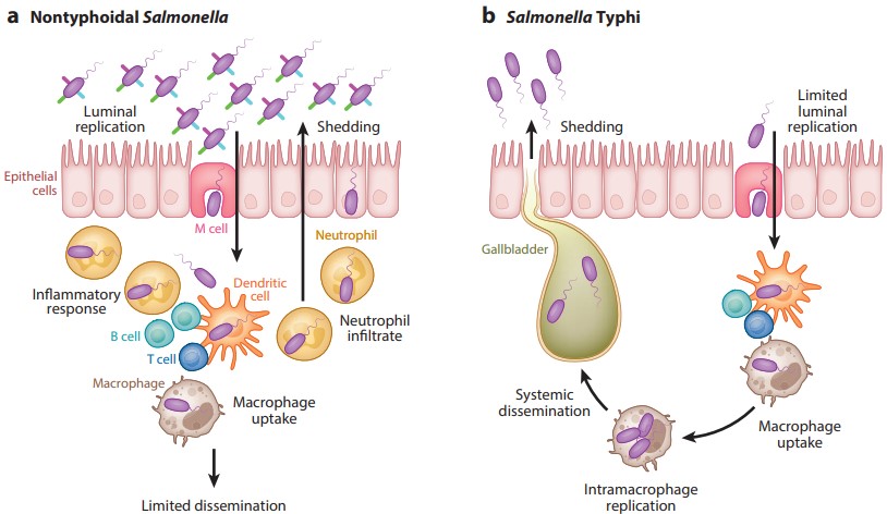 Figure 2. Typhoidal and nontyphoidal Salmonella infections
(Source: Dougan G, et al. 2014)
Although studies using Salmonella Typhimurium have been valuable in understanding Salmonella virulence, there are key differences between S. Typhi and S. Typhimurium, most notably the fact that Salmonella typhimurium causes predominantly gastroenteritis rather than systemic disease in humans. At the genomic level, although 89% of the genes are shared between the two serovars, nearly 500 genes are unique to Salmonella Typhimurium (strain LT2) and more than 600 genes are unique to Salmonella Typhi (strain CT18).
Among the genes specific to Salmonella Typhi are those encoding important virulence factors, including typhoid toxin and Vi antigen. In addition, both S. Typhi and S. Paratyphi A have a high proportion of pseudogenes (about 4%), which are associated with the restricted lifestyle of the host. In contrast, in Salmonella typhimurium, about 0.9% of the genes were pseudogenes. In addition, some studies have observed significant phenotypic differences between Salmonella Typhimurium and Salmonella serovars.
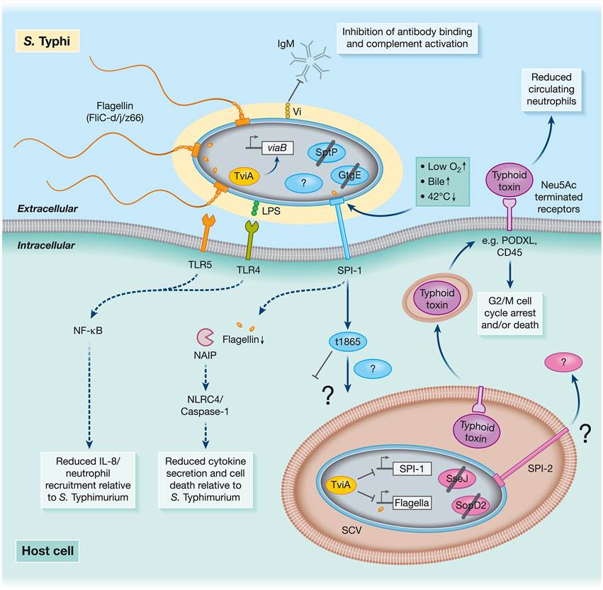 Figure 3. Molecular pathogenesis of Salmonella Typhi
(Source: Johnson R, et al. 2018)
The Vi Antigen
One of the main characteristics that distinguishes S. Typhi from NTS is that it produces a polysaccharide capsule called Vi antigen. Vi capsule can inhibit phagocytosis and enhance serum resistance, probably by protecting O-antigen from antibody attack. The genes encoding the Vi capsule include the viaB locus in Salmonella Pathogenicity Island (SPI)-7, which also encodes the type III secretion system (T3SS) effector SopE and the type IVB pilus. The viaB locus encodes genes involved in regulation (tviA), Vi biosynthesis (tviBCDE), export and retention of Vi on the bacterial cell surface.
TviA is a positive regulator that promotes the expression of the viaB locus, whereas it down-regulates the expression of the flagellar and the SPI-1 gene under high osmolarity conditions. Vi expression is down-regulated in the gut, flagella and SPI-1 play a role in epithelial cell invasion, whereas during systemic dissemination it is up-regulated in tissues, preventing the induction of antibody-mediated neutrophil responses. The inhibition of flagellin expression mediated by tviA leads to the limitation of NAIP recognition of flagellin, leading to reduced levels of pyroptosis and IL‐1β secretion by macrophages. Vi has been reported to bind cell surface prohibitin, thereby suppressing inflammation through MAPK signaling and IL-8 production. The decrease of IL-8 secretion mediated by TLR5 and TLR4 leads to the decrease of neutrophil influx. S. Paratyphi A lacks viaB, so it does not express Vi capsule. In contrast, S. Paratyphi A avoids antibody binding and antibody-mediated complement activation by forming long O-chains.
The Typhoid Toxin
S. Typhi is unique in that it expresses the typhoid toxin. The toxin is expressed when S. Typhi is located inside the cell and localized in the Salmonella-containing vacuole (SCV). Typhoid toxin is an atypical AB-type toxin consisting of two enzymatically active (A) subunits (CdtB and PltA) and a binding (B) subunit (PltB). CdtB is homologous to the A subunit of the cytotoxic distending toxin as well as to the DNase I family of proteins, whereas PltA, which has the ADP-ribosyl transferase-activity, and PltB, are similar to the subunits of the pertussis toxin. CdtB induces G2/M cell cycle arrest by damaging host cell DNA and inducing a DNA damage response through its DNase I-like activity. Homologs encoding typhoid toxin genes with slightly different sequences were found in Salmonella Paratyphi A and several NTS serovars, but not in Salmonella Typhimurium and Salmonella Enteritidis.
Typhoid toxin is secreted through SCV-derived vesicles and released extracellularly. After export, typhoid toxin is involved in the intoxication of infected and uninfected cells through autocrine and paracrine pathways. Typhoid toxin enters a range of target cells via PltB-mediated binding to glycans, primarily those terminated with N-acetylneuraminic acid (Neu5Ac). Glycosylated podocalyxin-like protein 1 (PODXL) on human epithelial cells and CD45 on immune cells, including macrophages, have been identified as typhoid toxin receptors. Neu5Ac is human-specific, which highlights the host limitations and adaptations of Salmonella Typhi. Notably, chronic typhoid carriers have been associated with gallbladder cancer. Therefore, it will be interesting to investigate whether typhoid toxins cause malignancy through their DNA damage activity.
The role of typhoid toxin in disease and pathogenesis remains unclear. After systemic administration of purified active typhoid toxin to mice, the mice develop lethargy, decreased circulating neutrophil counts, and neurological complications manifested by motor dysfunction, but not fever. Typhoid toxin is thought to be involved in the process of promoting chronic Salmonella Typhi infection, but its potential mechanism remains to be further studied. These studies suggest that typhoid toxin is the cause of symptom development and the transition from acute to chronic state during typhoid fever, and may be a target for reducing typhoid symptoms.
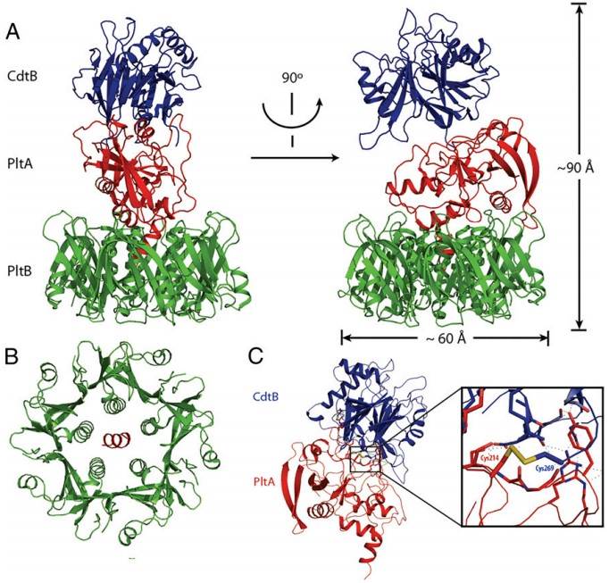 Figure 4. Atomic structure of typhoid toxin
(Source: Galán JE. 2016)
References
| Target | Cat. No. | Product Name | Size | Species Reactivity | Application | Detection Sample | |
| Salmonella | DEIABL37 | Chicken Salmonella Enteritidis IgY ELISA Kit | 96T | Quantitative | Serum, Plasma, other biological fluids | Inquiry | |
| DEIA2563 | Salmonella Antigen In Food ELISA Kit | 96T | Qualitative | Selected foods, production environmental samples | Inquiry | ||
| DEIA4172 | Salmonella IgG ELISA Kit | 96T | Human | Quantitative | Serum, plasma | Inquiry | |
| DEIA-WZ1001S | Salmonella abortusovis ELISA Kit | 96T | Sheep | Qualitative | Sheep Serum and Plasma | Inquiry |
| Target | Cat. No. | Product Name | Expression System | Tag/Conjugate | Application | |
| Salmonella | DAG-WT232 | Salmonella Typhi-O [BSA] | N/A | BSA | Immunoassays | Inquiry |
| DAG-WT525 | Inactivated Native Salmonella enteritidis Antigen | S. enteritidis | N/A | ELISA, LF | Inquiry | |
| S. typhi | DAGA-248 | Recombinant Salmonella typhi pagC Antigen [His] | E. coli | His | ELISA | Inquiry |
| DAGA-247 | Salmonella typhi pagC Antigen (full length) [His] | E. coli | His | N/A | Inquiry | |
| DAGA-3084 | Recombinant Salmonella typhi flag [His] | E. coli | His | EIA | Inquiry |
Loading ......
