
Inquiry Basket
 Loading ......
The Paramyxoviridae family contains human parainfluenza viruses (HPIVs) which belong to a large group of enveloped single-stranded RNA viruses. The Paramyxoviridae family represents a growing collection of viruses which contains human metapneumovirus along with mumps virus, measles virus, and respiratory syncytial virus. Genetic and antigenic analyses show that HPIVs consist of four main subtypes called HPIV-1 through HPIV-4. The human parainfluenza virus subtypes HPIV-1 and HPIV-3 belong to the genus Respirovirus while HPIV-2 and HPIV-4 fall under the genus Rubulavirus with both genera situated within the Paramyxovirinae subfamily. The Paramyxoviridae family members exhibit phenotypic differences with HPIVs through their neuraminidase activity presence and nucleocapsid thickness variation.
Table 1. Taxonomic classification of HPIV subtypes 1–4 within the Paramyxoviridae family
| Subfamily | Genus | Species |
| Paramyxovirinae | Respirovirus | Human parainfluenza virus 1 (HPIV-1) |
| Human parainfluenza virus 3 (HPIV-3) | ||
| Rubulavirus | Human parainfluenza virus 2 (HPIV-2) | |
| Human parainfluenza virus 4 (HPIV-4) | ||
| Mumps virus | ||
| Morbillivirus | Measle virus | |
| Megamyxovirus | Hendra virus | |
| Nipah virus | ||
| Pneumovirinae | Pneumovirus | Human respiratory syncytial virus |
| Metapneumovirus | Human metapneumovirus |
(Source: Pawełczyk M, et al. 2017)
The Paramyxovirus family consists of enveloped viruses that maintain their non-segmented negative-strand RNA genome mostly inside the nucleocapsid structure. The HPIV genome contains about 15,000 nucleotides which encode six structural proteins including nucleocapsid protein L and P proteins that secure viral RNA tightly as well as hemagglutinin-neuraminidase, fusion protein and membrane protein. The helical nucleocapsid containing viral RNA and N, P, and L proteins serves as the template for viral RNA synthesis instead of naked RNA. The N protein binds viral RNA and provides the template for transcription and replication by L and P proteins. Each N protein binds six nucleotides, so paramyxovirus genomes replicate most efficiently when the total number of nucleotides is a multiple of six, known as the "rule of six."
The outer layer of the virus contains spike structures made from HN and F proteins that enable host cell attachment and membrane fusion. HPIV-3 gains access to cells through direct fusion with the host cell membrane. The viral surface HN protein partners with the F protein to initiate fusion between the virus and cell membranes during entry. The interaction between the HN protein and the cell receptor activates a structural change in the F protein which allows it to perform fusion between the viral membrane and the host cell membrane. During the first stage of infection the viral negative-strand RNA genome coated with N protein undergoes transcription to generate a limited amount of viral mRNA. Then, the viral genome is replicated into a complementary positive-strand antigenome coated with N protein, which is subsequently used to generate new progeny genomes. Secondary transcription of the progeny genomes produces large amounts of viral mRNA, which are translated by cellular ribosomes into viral proteins. Newly synthesized negative-strand RNA is coated with N and P proteins to form new nucleocapsids, which can be used for further replication or assembled into new viruses for release.
Paramyxoviruses' attachment glycoprotein forms a tetramer structure that includes two dimers where every monomer binds to a single receptor molecule. The attachment glycoprotein experiences a conformational change that reveals its stalk region when it binds to its matching receptor which then activates the fusion glycoprotein to begin membrane fusion. The fusion glycoprotein belongs to the class I trimeric fusion proteins which contain abundant α-helical structures and need proteolytic cleavage from the precursor F0 for activation. Following its transport to the cell surface F0 undergoes endocytosis during which specific proteases like caspases and furin-like proteases cleave it into F1 and F2 subunits inside the endosome. The two subunits are linked by disulfide bonds and return to the cell membrane as an active fusion protein complex, executing the fusion of the viral and host cell membranes.
 Figure 1. Paramyxovirus attachment and fusion glycoprotein
(Source: Aguilar HC, et al. 2016)
Heptad repeat 1 (HR1) along with heptad repeat 2 (HR2) build the fusion core of the F protein trimer. The HR1 domain exists near the F1 subunit's N-terminus whereas the HR2 domain appears next to the C-terminus. An additional helical region known as HR3 is present upstream of the fusion peptide as well as within the F2 subunit. The HR1 and HR2 α-helical domains have a strong tendency to bind each other, forming an antiparallel coiled-coil bundle. The HR1 domain is located at the head of the fusion protein, is hydrophobic, and is shielded. In contrast, the HR2 domain is amphipathic (both hydrophilic and hydrophobic), exposed to the aqueous environment, and adopts a stalk-like conformation. Their binding releases energy that drives fusion between the virus and cell membranes. The HR3 domain does not bind HR1 or HR2 and may have other functions, but its role is not yet clear.
In the paramyxovirus-mediated virus-cell membrane fusion or cell-cell membrane fusion process, several key steps occur. Early steps include the attachment protein (such as HN, H, or G protein) binding to its corresponding cell surface receptor; followed by activation through conformational rearrangement of the attachment protein itself; then triggering large-scale conformational changes in the fusion protein, ultimately leading to membrane fusion. The late steps of membrane fusion include the formation and expansion of the fusion pore.
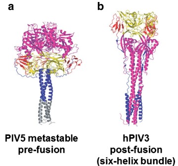 Figure 2. The pre- and post-fusion conformations of paramyxovirus F glycoproteins
(Source: Aguilar HC, et al. 2016)
Paramyxovirus attachment proteins (HN, H, or G) interact with the F protein in a way that is crucial for viral entry into host cells, but the timing of this interaction differs among viruses, leading to two main mechanisms: the "association model" and the "dissociation model."
The association model applies to paramyxoviruses whose HN glycoproteins bind sialic acid receptors, such as PIV5, HPIV3, and Newcastle disease virus. In these viruses, the HN protein binds the receptor first and then interacts with the F protein to activate it, promoting membrane fusion.
The dissociation model is relevant for viruses with H or G glycoproteins that attach to protein receptors including measles virus and other related viruses like canine distemper virus and both Nipah and Hendra viruses. During receptor binding the H or G protein first connects to the F protein and then loses this connection which leads to the activation of the F protein.
At least five models have been proposed for how the G protein triggers F protein activation: the sliding model, stalk-exposure model, safety-catch model, bi-dentate model, and oligomerization model. The sliding model suggests that after receptor binding, the attachment glycoprotein's head shifts laterally from a planar to a more staggered conformation, causing the stalk region to change from a compact four-helix bundle to a partially dissociated state. This releases the F protein from restraint, allowing it to activate freely. However, this model does not apply to all viruses, indicating diversity in F protein triggering mechanisms.
Membrane bilayer fusion is neither spontaneous nor simple. Activation of the F protein involves both proteolytic cleavage and interaction with the attachment protein, both of which are essential. F protein splits from precursor F0 into F1 and F2 molecules whereafter the fusion peptide at F1's N-terminus embeds itself into the target membrane leading HR1 and HR2 regions to adapt structurally to create a stable trimeric coiled-coil structure which facilitates membrane fusion. The F protein shifts through three stages during fusion moving from prefusion to fusion intermediate (PHI) to postfusion conformation while releasing energy to surpass membrane fusion's high energy barrier. F protein-mediated membrane fusion usually occurs under neutral pH conditions, but different viruses vary in entry mechanisms and environmental requirements; some may use endocytic pathways or depend on factors like calcium ions to complete infection. After activation, the fusion protein F inserts its fusion peptide into the target membrane, forming a transient intermediate state that can be inhibited by peptides derived from HR1 and HR2. Then, HR1 and HR2 refold and assemble into a six-helix bundle (6HB), bringing the fusion peptide and transmembrane region close together. The formation of the 6HB and its associated free energy changes are closely linked to the fusion of the viral and cellular membranes.
The F-GCNt prefusion structure and solF0 postfusion structure reveal how different folding intermediates of the F protein mediate membrane fusion and couple with F protein activation and fusion progression. Researchers have proposed a fusion model based on these findings. The HR2 helical structure first forms an "open stalk" conformation, breaking interactions at the base of the head and creating an early fusion intermediate, while HR1 remains in the prefusion conformation; peptide inhibition experiments support the existence of this stage. The opening of the HR2 stalk triggers conformational changes in other parts of the F protein. Notably, the F trimer may transiently dissociate, similar to conformational transitions proposed for other viral fusion proteins. Next, the DIII domain refolds, the HR1 coiled coil assembles, and the fusion peptide moves toward the target cell membrane, forming a pre-hairpin intermediate. After the fusion peptide moves away from the subunit interface, the DII domain can swing inward and form new contacts with the DI domain of a neighboring subunit, making the head structure more compact. The refolding of HR1 in DIII also exposes its core β-sheet structure, which, together with the inward swing of DII, promotes the formation of parallel β-strands between the HR2 linker region and the DIII core. This step likely precedes and initiates the final positioning of HR2. Ultimately, assembly of the 6HB completes the conformational change of the F protein and facilitates membrane fusion. The HN/H protein may promote F protein activation in a receptor-dependent manner by regulating the stability of the prefusion stalk of the F protein.
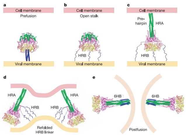 Figure 3. Model of F-mediated membrane fusion
(Source: Yin HS, et al. 2006)
References
| Target | Cat. No. | Product Name | Size | Species | Application | Detection Sample | |
| PIV | DEIA369 | Parainfluenza 1/2/3 IgG ELISA Kit | 96T | Human | Quantitative | Serum, plasma | Inquiry |
| DEIA370 | Parainfluenza Virus IgA ELISA Kit | 96T | Human | Quantitative | Serum, plasma | Inquiry | |
| DEIA371 | Parainfluenza 1/2/3 IgM ELISA Kit | 96T | Human | Quantitative | Serum, plasma | Inquiry | |
| DEIA1857 | Parainfluenza virus Antibody IgA ELISA Kit | 96T | Human | Qualitative | Serum, citrate plasma | Inquiry | |
| DEIA1948 | Parainfluenza 1/2/3 IgG ELISA Kit | 96T | Human | Qualitative | Human Serum or Plasma (citrate, heparin) | Inquiry | |
| DEIA1949 | Parainfluenza 1/2/3 IgM ELISA Kit | 96T | Human | Quantitative | Serum, plasma | Inquiry | |
| DEIA1950 | Parainfluenza 1/2/3 IgA ELISA Kit | 96T | Human | Qualitative | Human Serum or Plasma (citrate, heparin) | Inquiry | |
| DEIA1949NS | Human anti-parainfluenza virus(PIV) antibody(IgM) ELISA Kit | 96T | Human | Quantitative | Serum | Inquiry |
Loading ......
