
Inquiry Basket
 Loading ......
Hepatitis B virus (HBV) infection is a significant global health issue, affecting millions of people and causing a high number of deaths each year. Mutations in the hepatitis B surface antigen (HBsAg) have been identified, primarily involving single amino acid substitutions or nucleotide deletions. These mutations are commonly found in the S region but also occur in the pre-S1 and pre-S2 regions of HBsAg. Single amino acid substitutions in the crucial hydrophilic region known as the "a" determinant can lead to immune escape, failing HBV vaccination and HBsAg detection. Deletions in the pre-S1 or pre-S2 regions have been associated with the development of hepatocellular carcinoma (HCC). Therefore, it is necessary to pay attention to HBsAg mutants and their biological and clinical significance.
Chronic HBV infection follows a complex and nonlinear natural history consisting of five distinct phases. These phases include HBeAg-positive chronic HBV infection, HBeAg-positive chronic hepatitis B (CHB), HBeAg-negative chronic HBV infection, HBeAg-negative active CHB, and the HBsAg-negative phase. In the HBsAg-negative phase, patients may still experience viral reactivation under conditions of severe immunosuppression. The current management of chronic HBV infection involves two therapeutic approaches: pegylated interferon α (PEG-IFNα) and nucleos(t)ide analogues (NAs). The primary treatment goal is to achieve long-term improvement by persistently inhibiting HBV replication.
HBV is the prototype member of the Hepadnaviridae family, which includes similar avian and mammalian viruses. The infectious HBV particle, known as a "Dane particle," is composed of a nucleocapsid core and a glycolipid envelope. The nucleocapsid contains partially double-stranded circular DNA, endogenous polymerase, and an icosahedral capsid. The envelope consists of a lipid bilayer with three distinct surface proteins.
HBV replicates through an RNA intermediate anti-genome sequence, utilizing its own reverse transcriptase (RT) enzyme. However, due to the lack of proofreading function in HBV-RT, errors occur during replication, similar to retroviruses and other RNA viruses. These error-prone conditions, combined with persistent and long-term infections and different selective pressures, have led to the emergence of HBV variants that can evade diagnostics, prophylactic measures, and therapeutic interventions.
The HBV genome consists of four partially overlapping open reading frames (ORFs) encoding specific proteins. The surface (preS/S) gene encodes surface proteins (HBsAg), including the small (S), middle (M), and large (L) forms. The core (preC/C) gene produces the core protein (HBcAg) and the e antigen (HBeAg). The polymerase (P) gene encodes the reverse transcriptase enzyme, and the X gene codes for a transcriptional transactivator protein.
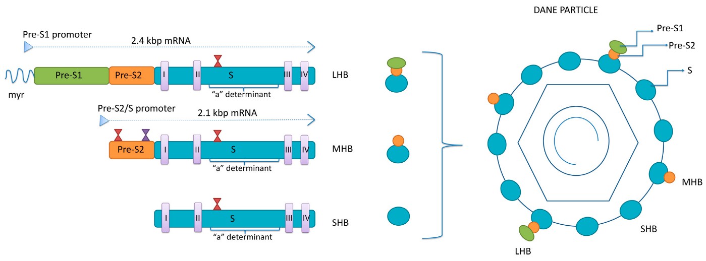 Figure 1. The structure of HBsAg isoforms.
(Source: Lazarevic, I. et al., 2024)
HBV genotypes are classified based on nucleotide differences within the genome sequence. Currently, nine well-defined genotypes and numerous sub-genotypes have been identified. These genotypes exhibit significant nucleotide differences, with more than 8% divergence for genotypes and 4-8% divergence for sub-genotypes. Over 40 related sub-genotypes have been determined thus far.
Earlier classification systems correlated HBV genotypes with four major serological subtypes of HBsAg: adw, adr, ayw, and ayr. Genotypes A, B, F, G, and H typically have the adw subtype, while genotype C has adr, and genotypes D and E have ayw.
HBsAg mutants refer to variants of the hepatitis B surface antigen (HBsAg) protein that have specific genetic changes or mutations. These mutations can occur naturally or as a result of selective pressures, such as immune responses or antiviral treatment. The clinical significance of HBsAg mutants lies in their potential to affect the diagnosis, prognosis, and management of hepatitis B virus (HBV) infection.
The first strategy is to increase antibodies against different sites on HBsAg. Currently, the mainstream fully automated chemiluminescent HBsAg detection kits mostly use a double-antibody sandwich detection method, and more than two monoclonal antibodies are often selected for the HBsAg epitope. New detection kits, such as the LIAISON XL Murex HBsAg detection kit, use 7 monoclonal antibodies targeting different epitopes of HBsAg, including the "a" epitope, transmembrane region, and intramembrane region. Such an antibody design effectively ensures the sensitivity of HBsAg detection and avoids missed detection due to "a" epitope mutation.
Another strategy is to open the spatial structure of HBsAg to form a linear structure that fully exposes the binding sites for antibodies and antigens. The binding of antibodies to epitopes requires full exposure of the epitopes, especially those targeting transmembrane and intramembrane sites, which requires specific reagents to dissociate HBsAg from the membrane. Different dissociation strategies can be used, such as using strong denaturants to allow monoclonal antibodies targeting different epitopes to fully bind and react with the "a" epitope, transmembrane region, and intramembrane region of HBsAg.
References
| Target | Cat. No. | Product Name | Size | Species Reactivity | Application | Detection Sample | |
| HBsAg | DEIA001 | Hepatitis B Surface Antigen ELISA Kit | 96T | Human | Qualitative | blood | Inquiry |
| DEIA060 | Antibody to HBsAg ELISA Kit | 96T | Human | Quantitative | Serum, plasma | Inquiry | |
| IVDEIA001 | Human Hepatitis B surface antigen (HBsAg) ELISA Kit | 96T | Human | Qualitative | Serum, plasma | Inquiry | |
| DEIASL263 | Mouse Anti-HBsAg IgG ELISA Kit | 96T | Quantitative | Srerum | Inquiry | ||
| DEIASL271 | Rabbit Anti-HBsAg IgG ELISA Kit | 96T | Quantitative | Srerum | Inquiry |
Loading ......
