











Inquiry Basket
 Loading ......
CD constitute an internationally recognized nomenclature for identifying cell-surface molecules on human and other vertebrate leukocytes. In short, CD molecules are surface proteins, each given a number, that are important for signal transduction, cell–cell adhesion and communication with the microenvironment. The CD system has revolutionized the field of immunology, by making it possible, for instance, to unambiguously identify, isolate and study specific subsets of immune cells, thanks to monoclonal antibodies that recognize the specific CDs that those cells express, for instance, distinguishing T cells and their major subtypes according to their expression of CD3, CD4 and CD8. CD markers are also of major importance in oncology, where aberrant expression of certain CDs on tumor cells may identify potential targets for biomarkers to facilitate early detection, staging and prognostication and even, more importantly, potential "docking sites" for targeted therapy, in particular antibody–drug conjugates and CAR-T treatments. Many more family members are being identified and defined all the time, and the complexity of the functional interplay and potential role of CD systems in disease is beginning to be uncovered with ever greater depth and detail.
T cells co-express the T cell receptor (TCR) and CD3 on their surface, which together mediate antigen recognition and activation signaling.
CD4 and CD8 respectively define helper T cells (Th) and cytotoxic T cells (Tc).
CD28 and CTLA-4 (also known as CD152) serve as the principal costimulatory receptors, driving T cell activation and inhibition.
A vast and mature research and clinical ecosystem has developed around CD markers, spanning from basic research tools to therapeutic drugs.
Diagnostic and research reagents
Diagnostic and research reagents are centered on specific monoclonal antibodies: fluorescent or isotope-labeled anti-CD antibodies are widely used in flow cytometry, IHC, and mass cytometry for cell sorting, phenotyping, and functional studies.


Technologies have evolved from conventional flow cytometry to high-dimensional mass cytometry, and now to single-cell sequencing and spatial omics platforms—each iteration revealing CD marker complexity at single-cell and tissue-spatial resolution.
Therapeutic drug development
CD molecules are among the most successful immunotherapy targets—this includes monoclonal antibodies such as rituximab targeting CD20 and cell therapies like CAR-T cells against CD19, which have achieved remarkable success in treating hematologic malignancies.

 Figure 1. Scatter plots showing spheroid area under the curve with isolated CD4+ (left) or CD8+ (center) T-cell fractions, or unseparated T cells (right)
(Source: Anderson VE, et al. 2023)
 Figure 2. Representative bioluminescence image of tumour burden in C57Bl/6 mice with ID8 wild-type versus ID8(ΔCd24a) tumours
(Source: Barkal AA, et al. 2019)

Han
Positive multi-tissue
In addition to Immunohistochemical analysis of paraffin-embedded human tonsillitis tissue slide using CABT-23576MH (CD63 antibody) at dilution of 1:700. Also tested positive in human tonsillitis tissue, human malignant melanoma tissue, human colon cancer tissue.
Sam
Worth buying!
An antibody worth buying! Immunohistochemical analysis of paraffin-embedded human lymphoma using CD7 antibody at dilution of 1:50 (under 10x lens).
Nazer
Applies to FC
Intracellular flow cytometric analysis of 4% paraformaldehyde fixed 90% methanol permeabilized Jurkat (Human T cell leukemia T lymphocyte) cells labelling CD7 with purified Anti-CD7 monoclonal antibody at 1/100 dilution.
Sam
Worth buying
Jurkat cells were subjected to SDS PAGE followed by western blot with this CD7 antibody at dilution of 1:1000 incubated at room temperature for 1.5 hours.
Single-omics data can only reveal one aspect of cellular state. The future trend is multi-layer information integration. For example, one can integrate CITE-seq (transcriptome + surface proteome), spatial omics and epigenomic information. By integrating these modalities, one can construct a more complete 3D cellular atlas and also understand the intricate wiring logic between gene regulation, protein activity and spatial context under specific physiological or pathological states.
Spatial omics is emerging as a "new frontier" in tumor immunology. Mapping CD markers in space can address important questions: In patients who respond to immune checkpoint inhibitors, how are CD8+ T cells and PD-L1+ cells co-localized in the tumor? How are different TAM subsets (CD68, CD163, etc.) distributed in different tumor regions? Are exhausted T cells (CD39, TIM-3, LAG-3, etc.) localized in certain "immunosuppressive niches"? Solving the above questions is critical to understand the mechanisms of resistance and to design the next generation of combination therapies.
High-throughput, high-dimensional technologies generate massive and complex datasets which are beyond human capacity for interpretation. AI and ML algorithms are required to make sense of these data. Computational biologists are inventing and applying novel algorithms to automatically identify cell populations, discover rare disease-associated subtypes and select the most predictive combination of CD markers from high-dimensional data. For example, ML models can predict a patient's response to immune checkpoint inhibitors from pre-treatment spatial omics profiles of their tumor, to enable truly personalized immunotherapy.

CD319 is a glycoprotein expressed on various immune cells, with especially high levels on NK cells and plasma cells. On NK cells, it functions primarily as an activating receptor that enhances tumor cell killing, though in certain contexts it may also transmit inhibitory signals. In oncology, CD319 has been validated as a therapeutic target.
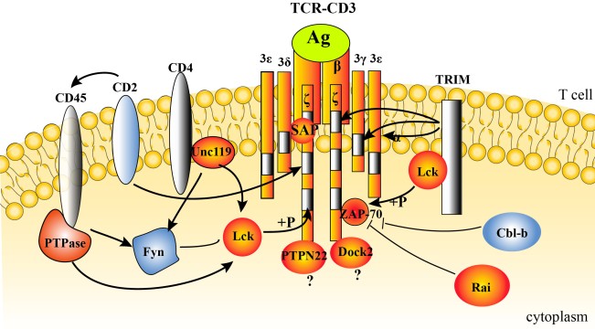
CD247 is an integral part of T cell signal transduction. Upon recognition of an antigen, ITAMs of CD247 become phosphorylated, initiating a series of downstream signals which lead to T cell activation, proliferation and effector function. As a result, expression levels of CD247 on peripheral blood or tumor-infiltrating T cells have become a critical index for evaluation of the immune status of the patients and for prediction of response to immunotherapies and clinical prognosis.
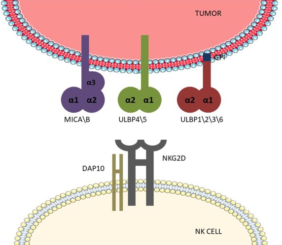
CD314 is an activating receptor with a wide expression on NK cells, CD8+ T cells and γδ T cells. NKG2D is the so-called "stress sensor" of the immune system because it does not recognize classical MHC molecules but rather ligands that are only weakly expressed on healthy cells but become upregulated under stress conditions, such as infection, DNA damage, and malignant transformation. These ligands include MICA/B and ULBPs.
References
Loading ......
