

Inquiry Basket
 Loading ......
The immune system is a complex network of cells, tissues, and organs that work together to protect the body from harmful pathogens, such as bacteria and viruses. To understand the immune system and how it fights against diseases, it is essential to study the molecules involved in immune cell recognition and activation. One of the most significant groups of molecules in this regard is CD antigens.
CD antigens, also known as clusters of differentiation antigens, are a group of cell surface molecules expressed on various cell types. They are used as markers to identify and classify cells, particularly those of the immune system. CD antigens are categorized as type I transmembrane proteins, which means that they have a single transmembrane domain and an extracellular domain that interacts with other molecules, such as ligands or antibodies. Over 300 CD antigens have been identified in humans, and they are classified based on their structure and function.
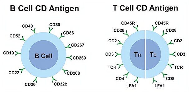
The current naming system for CD antigens was established in 1982 at the 1st International Workshop and Conference on Human Leukocyte Differentiation Antigens (HLDA). The HLDA conferences will continue to be held to maintain and develop the list of known CD markers. Under this system, well-characterized antigens are assigned an arbitrary number, such as CD1 and CD2, while molecules recognized by just one monoclonal antibody are given the provisional designation "CDw", such as CDw168. Lowercase letters are added after the assigned number to indicate larger molecules that share a common chain (e.g., CD1a, CD1b, CD1c, CD1d, or CD1e).
CD molecules have a wide range of functions that range from cell surface receptors to adhesion molecules, and they do not belong to any specific class physiologically. While initially used only for human leukocytes, the CD molecule naming system now includes different species and other cell types. As of April 2016, the list of human CD antigens has expanded to include up to CD371.
When a specific antigen is present or absent on the surface of a cell population, it is denoted by a "+" or a "-" respectively. For example, a "CD34+, CD31-" cell is one that expresses CD34, but not CD31. Overall variability in CD expression is marked as hi, mid, or low (alternatively bright, mid, or dim), such as CD62 low for effector memory T-cells. CD antigen expression profiles can be used to identify, isolate, and phenotypically characterize cell types based on their function in various immune processes.
CD antigens play critical roles in various aspects of immunology, including lymphocyte development, activation, and differentiation. For example, CD19 is a B cell-specific molecule involved in B cell development and activation. Defects in CD19 expression or function can lead to immunodeficiency disorders, such as common variable immunodeficiency.
CD antigens are also involved in the recognition and elimination of foreign substances, such as pathogens and tumors. For example, CD8 is a T cell-specific molecule involved in virus-infected and tumor cell recognition and killing. CD8 T cells recognize antigens presented on infected or cancerous cells by MHC class I molecules and then eliminate the cells through cytotoxic molecules.
In addition to their role in immunology, CD antigens have been implicated in the pathogenesis of various diseases, including cancer. For example, CD47 is a cell surface protein overexpressed on many types of cancer cells, and it plays a critical role in immune evasion by inhibiting phagocytosis by macrophages. Therapeutic targeting of CD47 has been shown to enhance anti-tumor immunity and improve cancer immunotherapy efficacy.
CD antigens are used to identify populations and subpopulations of immune system cells through the use of antibodies to detect markers on the surface of cells. They are commonly used for immunophenotyping and can be expressed only at certain stages of development or under particular conditions. Some CD antigens are unique to a specific cell lineage. For example, CD20 is exclusively expressed on lymphocytes of the B-cell lineage. The following table shows CD markers used for immune typing:
| Type of cell | CD markers |
| Stem cells | CD34+, CD31-, CD117 |
| all leukocyte groups | CD45+ |
| Granulocyte | CD45+, CD11b, CD15+, CD24+, CD114+, CD182+ |
| Monocyte | CD4, CD45+, CD14+, CD114+, CD11a, CD11b, CD91+, CD16+ |
| T lymphocyte | CD45+, CD3+ |
| T helper cell | CD45+, CD3+, CD4+ |
| T regulatory cell | CD4, CD25, FOXP3 |
| Cytotoxic T cell | CD45+, CD3+, CD8+ |
| B lymphocyte | CD45+, CD19+, CD20+, CD24+, CD38, CD22 |
| Thrombocyte | CD45+, CD61+ |
| Natural killer cell | CD16+, CD56+, CD3-, CD31, CD30, CD38 |
At Creative Diagnostics, we understand the importance of CD antigens in immunology research, and we are committed to providing our customers with the highest quality primary antibodies for the detection of these molecules. Our human CD antigen guide provides a comprehensive list of over 300 CD antigens, including their names, descriptions, and functions. You can contact us for a complete list.
Our antibodies are rigorously tested for specificity and sensitivity, and we offer a range of formats, including monoclonal and polyclonal antibodies, as well as conjugated and unconjugated antibodies. We also offer a range of related resources, such as protocols for using antibodies, a tool for designing multicolor flow cytometry experiments, and a glossary of terms related to CD antigens and immunology.
Loading ......
