
Inquiry Basket
 Loading ......
CD68, also known as colony-stimulating factor 1 receptor-associated protein (CSF1R-associated protein) or macrophage antigen, is a biomarker with extensive use in immunological and pathological research. A transmembrane glycoprotein abundantly expressed by cells of the monocyte and macrophage lineage, CD68 has been used for decades as a reliable marker for identification and quantification of macrophages in tissues.
Browse all CD68 related products
CD68 protein is a member of the lysosomal/endosome-associated membrane protein (LAMP) family. The human gene for this protein is located on chromosome 17. CD68 is a highly glycosylated type I transmembrane protein. CD68 is most abundantly located on the membranes of intracellular organelles such as lysosomes and endosomes. It is less commonly located on the surface of the cell. The extracellular domain of CD68 is heavily glycosylated and can interact with selectins or specific tissue lectins. CD68 is functionally and evolutionarily related to other gene or protein families such as hematopoietically expressed mucin like molecules (e.g. CD43, leukosialin and the hematopoietic stem cell antigen CD34).
CD68 gene expression is strictly controlled by several transcription factors including PU.1, c-Jun, Ets family transcription factors, and interferon regulatory factors, which collectively determine its highest and specific expression in myeloid cells, in particular, macrophages. CD68 is also found in a variety of other cells types including dendritic cells, osteoclasts, and some fibroblasts, albeit, usually during specific physiological or pathological conditions. CD68 has been shown to bind a number of ligands, such as oxidized low-density lipoprotein (oxLDL), phosphatidylserine, and apoptotic cells, and is thought to have scavenger receptor activity and a role in lipid metabolism and clearance of apoptotic cells.
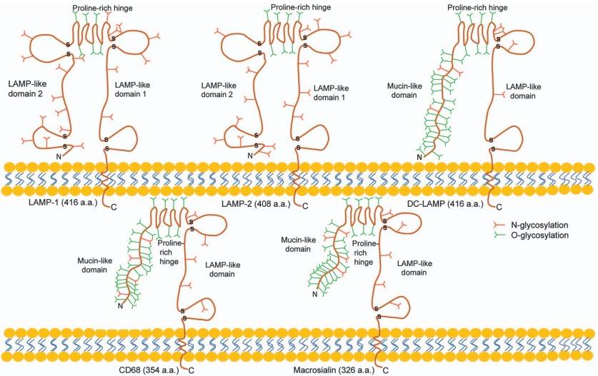 Figure 1. A scheme shows the general features of the domain organization of murine macrosialin and human proteins LAMP-1, LAMP-2, DC-LAMP, and CD68 and depicts the principal differences between the family members.
(Source: Chistiakov DA, et al. 2017)
In most histopathological and immunological contexts, "CD68 positive" is considered to be a definitive marker for the presence of macrophages. By IHC staining of tissue sections, CD68 positive cells can be easily visualized and enumerated, and therefore the degree of macrophage infiltration can be determined. For this reason, CD68 is taken to be a pan-marker of the total infiltrating macrophage population.
However, CD68 has inherent limitations as a macrophage marker. Macrophages are highly plastic and can polarize into functionally distinct subtypes in response to signals from the microenvironment, most classically the pro-inflammatory M1 macrophages and the anti-inflammatory/pro-tumoral M2 macrophages. Studies have shown that CD68 itself cannot effectively distinguish between these two functionally divergent macrophage subsets. Both M1 and M2 macrophages are typically CD68-positive. This characteristic complicates the use of CD68-positive cell counts alone to determine their functional and prognostic impact in specific diseases, such as cancer, and is a key reason for inconsistent findings across many clinical studies.
 Figure 2. Typical expression pattern of the macrophage markers CD68, CD11c, CD163 and MRC1 in a lymph node
(Source: Wehrhan F, et al. 2024)
In the field of cancer research, CD68+ macrophages are often denoted as tumor-associated macrophages (TAMs). Tumor infiltration density of TAMs is often associated with tumor progression and poor prognosis. For instance, in breast cancer, not only is the overall density of CD68+ TAMs increased, the CD68+/CD206+ subpopulation (CD206 is a marker of M2 macrophages) is significantly associated with larger tumor diameter, higher lymph node metastasis rate and lower survival rate. CD68 expression was positively correlated with the other macrophage markers (CD163 and CD11b) and immune checkpoint molecules (PD-1 and TIM-3) in glioma, which indicated its involvement in the establishment of an immunosuppressive microenvironment.
CD68-positive macrophages are also involved in other inflammatory and autoimmune diseases. In renal tissues of lupus nephritis (LN) patients, infiltration of CD68-positive cells is significantly enhanced and it positively correlates with the degree of injury both in glomeruli and renal tubulointerstitium, suggesting an active involvement of macrophages in pathogenic processes in the kidney.
As a member of the scavenger receptor family, one of CD68's core functions is its involvement in lipid metabolism and phagocytosis. CD68 contributes to the phagocytic activity of macrophages through its roles in intracellular lysosomal metabolism, as well as in extracellular interactions between cells or between cells and pathogens. In macrophages, CD68 molecules bind to tissue and organ-specific lectins or selectins, enabling macrophages to target specific entities or directing macrophage subsets to home to specific sites. Furthermore, the rapid recycling of CD68 from endosomes and lysosomes to the cell membrane allows macrophages to move along the surfaces of selectin-expressing cells or other substrates.
Multiple studies have confirmed that CD68 can bind to and internalize OxLDL. This function is particularly significant in the pathogenesis of atherosclerosis, where macrophages ingest OxLDL and transform into foam cells, thereby promoting plaque formation. CD68 also participates in the phagocytosis of apoptotic cells, cellular debris, and certain pathogens such as bacteria. Through this process, macrophages not only eliminate potentially harmful substances but may also participate in antigen processing and presentation, thereby initiating adaptive immune responses.
The tumor microenvironment is composed of various cells, including leukocytes, fibroblasts, vascular endothelial cells, and immune cells, with the latter being a critical component. These cells interact with tumor cells and influence tumor initiation, growth, and metastasis. Within this microenvironment, macrophages are termed tumor-associated macrophages (TAMs) and represent one of the most abundant immune cell populations.
Activated macrophages are broadly categorized into two main types. When stimulated by Th1-type cytokines such as IFN-γ or bacterial products, macrophages are classically activated into M1-type macrophages. M1 macrophages are considered inhibitory; they express CD68 and HLA-DR, and exert cytotoxic effects through antigen presentation and phagocytosis of tumor cells. They suppress tumor growth by generating reactive oxygen species (ROS), reactive nitrogen intermediates, and tumor necrosis factor.
On the other hand, when the microenvironment is rich in cytokines such as IL-4, IL-13, IL-10, glucocorticoids, or transforming growth factor, macrophages are alternatively activated and polarized into M2-type macrophages. M2 macrophages are also CD68+ with other markers such as CD163. M2 macrophages are major players in Th2-type immune responses. M2 TAMs promote tumor progression by promoting angiogenesis, inducing immunosuppression, and tumor cell survival, proliferation, apoptosis resistance, infiltration, invasion, and metastasis. As a result, M2 TAMs are linked to greater tumor growth, therapy resistance, and poor prognosis. Recent studies have found that M2 TAMs are found in abundance in tumor tissues. The cytokines they produce and release have important roles in creating an immunosuppressive TME.
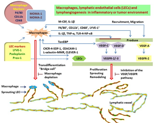 Figure 3. The schema shows macrophage-induced lymphangiogenesis
(Source: Ji RC. 2012)
CD68 immunohistochemistry is considered the "gold standard" method for assessing the overall infiltration level of TAMs in solid tumors. By staining and quantitatively analyzing tumor tissue sections for CD68—such as by calculating the number of positive cells per unit area—researchers can obtain key information regarding TAM abundance. However, since CD68 cannot distinguish between functional subtypes of TAMs, studies examining the relationship between CD68-positive TAM density and patient prognosis often yield conflicting conclusions. Many studies have shown that high density of CD68-positive TAMs is significantly associated with poorer overall survival (OS) and disease-free survival (DFS) in various solid tumors, such as certain sarcomas, non-small cell lung cancer, breast cancer, and hepatocellular carcinoma. This is often interpreted as TAMs in these tumors predominantly exhibiting a pro-tumor M2-like phenotype, promoting angiogenesis, suppressing anti-tumor immunity, and facilitating metastasis. In contrast, other studies have found no significant association between CD68-positive cell density and patient prognosis, or have even observed a correlation with better outcomes in some cancer types. This favorable association may occur in tumor microenvironments where M1-like macrophages dominate, effectively presenting tumor antigens and killing tumor cells. This contradiction in prognostic value highlights the limitation of relying solely on CD68 for outcome prediction. Future studies must incorporate additional markers—such as CD163 or CD206 for M2-like polarization, and iNOS or HLA-DR for M1-like polarization—to enable more refined analysis of TAM subsets.
Additionally, the role of TAMs in modulating treatment responses has attracted significant attention. Emerging research has revealed potential links between CD68 expression and therapeutic efficacy. Some studies indicate that high CD68 expression may be associated with longer survival in patients receiving immune checkpoint inhibitor therapy. This suggests that a macrophage-rich, "inflamed" tumor microenvironment might be more sensitive to certain immunotherapies, though this still requires validation through larger prospective clinical studies.
References
| Target | Cat. No. | Product Name | Size | Species | Application | Detection Sample | |
| CD68 | DEIA-BJ443 | Human CD68(Macrosialin) ELISA Kit | 96T | Human | Quantitative | Serum, plasma, tissue homogenates, other biological fluids | Inquiry |
| Target | Cat. No. | Product Name | Expression System | Tag/Conjugate | Application | |
| CD68 | DAG-KO028 | CD68 Knockout Cell Lysate | WB | Inquiry |
Loading ......
